

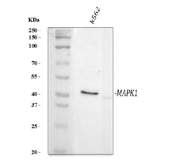
Figure 1. Western blot analysis of MAP kinase,activated using anti-MAP kinase,activated antibody (MA1055). Electrophoresis was performed on a 5-20% SDS-PAGE gel at 70V (Stacking gel) / 90V (Resolving gel) for 2-3 hours. The sample well of each lane was loaded with 30 ug of sample under reducing conditions. Lane 1: human K562 whole cell lysates. After electrophoresis, proteins were transferred to a nitrocellulose membrane at 150 mA for 50-90 minutes. Blocked the membrane with 5% non-fat milk/TBS for 1.5 hour at RT. The membrane was incubated with mouse anti-MAP kinase,activated antigen affinity purified monoclonal antibody (Catalog # MA1055) at 0.5 microg/mL overnight at 4°C, then washed with TBS-0.1%Tween 3 times with 5 minutes each and probed with a goat anti-mouse IgG-HRP secondary antibody at a dilution of 1:10000 for 1.5 hour at RT. The signal is developed using an Enhanced Chemiluminescent detection (ECL) kit (Catalog # EK1001) with Tanon 5200 system. A specific band was detected for MAP kinase,activated at approximately 42 kDa. The expected band size for MAP kinase,activated is at 41 kDa.
Figure 1. Western blot analysis of MAP kinase,activated using anti-MAP kinase,activated antibody (MA1055). Electrophoresis was performed on a 5-20% SDS-PAGE gel at 70V (Stacking gel) / 90V (Resolving gel) for 2-3 hours. The sample well of each lane was loaded with 30 ug of sample under reducing conditions. Lane 1: human K562 whole cell lysates. After electrophoresis, proteins were transferred to a nitrocellulose membrane at 150 mA for 50-90 minutes. Blocked the membrane with 5% non-fat milk/TBS for 1.5 hour at RT. The membrane was incubated with mouse anti-MAP kinase,activated antigen affinity purified monoclonal antibody (Catalog # MA1055) at 0.5 microg/mL overnight at 4°C, then washed with TBS-0.1%Tween 3 times with 5 minutes each and probed with a goat anti-mouse IgG-HRP secondary antibody at a dilution of 1:10000 for 1.5 hour at RT. The signal is developed using an Enhanced Chemiluminescent detection (ECL) kit (Catalog # EK1001) with Tanon 5200 system. A specific band was detected for MAP kinase,activated at approximately 42 kDa. The expected band size for MAP kinase,activated is at 41 kDa.
Anti-Phospho-MAP Kinase, Activated(Diphosphorylated ERK-1&2) Antibody (Monoclonal, MAPK-YT)
MA1055
ApplicationsWestern Blot, ImmunoCytoChemistry, ImmunoHistoChemistry
Product group Antibodies
ReactivityBovine, Human, Monkey, Mouse, Rat, Yeast
TargetMAPK1
Overview
- SupplierBoster Bio
- Product NameAnti-Phospho-MAP Kinase, Activated(Diphosphorylated ERK-1&2) Antibody (Monoclonal, MAPK-YT)
- Delivery Days Customer9
- Application Supplier NoteOther applications have not been tested. Optimal dilutions should be determined by end users.
- ApplicationsWestern Blot, ImmunoCytoChemistry, ImmunoHistoChemistry
- Applications SupplierIHP, ICC, WB, IHC
- CertificationResearch Use Only
- ClonalityMonoclonal
- Clone IDMAPK-YT
- Concentration100 ug/ml
- Gene ID5594
- Target nameMAPK1
- Target descriptionmitogen-activated protein kinase 1
- Target synonymsERK, ERK-2, ERK2, ERT1, MAPK2, NS13, P42MAPK, PRKM1, PRKM2, p38, p40, p41, p41mapk, p42-MAPK, mitogen-activated protein kinase 1, MAP kinase 1, MAP kinase 2, MAPK 2, extracellular signal-regulated kinase 2, mitogen-activated protein kinase 2, protein tyrosine kinase ERK2
- HostMouse
- IsotypeIgG1
- Protein IDP21708
- Protein NameMitogen-activated protein kinase 3
- Scientific DescriptionBoster Bio Anti-Phospho-MAP Kinase, Activated (Diphosphorylated ERK-1&2) Mapk3 Antibody (Monoclonal, MAPK-YT) catalog # MA1055. Tested in IHC, ICC, WB applications. This antibody reacts with Human, Mouse, Rat, Yeast.
- ReactivityBovine, Human, Monkey, Mouse, Rat, Yeast
- Storage Instruction-20°C,2°C to 8°C
- UNSPSC12352203
References
- Lu J, Yang Y, Varga E, et al. Molecular Mechanisms Associated with Protecting IEC-6 Cells from Acrylamide-Induced Tight Junction Damage by Ganoderma atrum Polysaccharide. Mol Nutr Food Res. 2023,67(6):e2200774. doi: 10.1002/mnfr.202200774Read this paper
- Han M, Wang X, Wang J, et al. Ameliorative effects of epigallocatechin-3-gallate nanoparticles on 2,4-dinitrochlorobenzene induced atopic dermatitis: A potential mechanism of inflammation-related necroptosis. Front Nutr. 2022,9:953646. doi: 10.3389/fnut.2022.953646Read this paper
- Wu HB, Wang ZW, Shi F, et al. Avβ3 single-stranded DNA aptamer attenuates vascular restenosis via Ras-PI3K/MAPK pathway in rats after percutaneous transluminal coronary angioplasty. Artif Organs. 2020,44(6):611-619. doi: 10.1111/aor.13622Read this paper
- Jiang A, Zhang Y, Zhang X, et al. Morin alleviates LPS-induced mastitis by inhibiting the PI3K/AKT, MAPK, NF-κB and NLRP3 signaling pathway and protecting the integrity of blood-milk barrier. Int Immunopharmacol. 2020,78:105972. doi: 10.1016/j.intimp.2019.105972Read this paper
Datasheet
MSDS
Related products
Product group Antibodies
MAPK1 Polyclonal AntibodyCAC14542
ApplicationsImmunoFluorescence, Western Blot, ELISA, ImmunoHistoChemistry
ReactivityRat
TargetMAPK1
- SizePrice
Product group Antibodies
Anti-ERK1 / ERK2 Antibody144-64587
ApplicationsImmunoFluorescence, Western Blot, ImmunoHistoChemistry
ReactivityHuman, Mouse, Rat
TargetMAPK1
- SizePrice
Product group Antibodies
References
ApplicationsFlow Cytometry, ImmunoFluorescence, Western Blot, ELISA, ImmunoCytoChemistry, ImmunoHistoChemistry, ImmunoHistoChemistry Frozen, ImmunoHistoChemistry Paraffin
ReactivityBovine, Canine, Chicken, Equine, Guinea Pig, Human, Mouse, Porcine, Rabbit, Rat
TargetMAPK1
- SizePrice
Product group Antibodies

ApplicationsImmunoFluorescence, Western Blot, ELISA
ReactivityBovine, Canine, Human, Mouse, Rat
TargetMAPK1
- SizePrice
Product group Antibodies
ERK2 antibody [N1], N-termGTX104613
ApplicationsImmunoFluorescence, Western Blot, ImmunoCytoChemistry, ImmunoHistoChemistry, ImmunoHistoChemistry Paraffin
ReactivityCanine, Feline, Human
TargetMAPK1
- SizePrice
Product group Antibodies
Anti-ERK1/2 AntibodyA29693
ApplicationsImmunoFluorescence, Western Blot, ImmunoHistoChemistry
ReactivityHuman, Mouse, Rat
- SizePrice

Product group Antibodies
References
ApplicationsFlow Cytometry, ImmunoFluorescence, ImmunoPrecipitation, Western Blot, ImmunoCytoChemistry, ImmunoHistoChemistry
ReactivityHuman, Mouse, Rat
TargetMAPK1
- SizePrice
Product group Antibodies
MAPK3/MAPK1 AntibodyCSB-PA002419
ApplicationsImmunoFluorescence, Western Blot, ELISA
ReactivityHuman, Mouse, Rat
TargetMAPK1
- SizePrice