


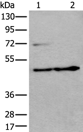
Figure 1. Western blot analysis of PKA R2/PKR2/PRKAR2A using anti-PKA R2/PKR2/PRKAR2A antibody (A03911-2). Electrophoresis was performed on a 5-20% SDS-PAGE gel at 70V (Stacking gel) / 90V (Resolving gel) for 2-3 hours. The sample well of each lane was loaded with 30 ug of sample under reducing conditions. Lane 1: human K562 whole cell lysates, Lane 2: human Jurkat whole cell lysates, Lane 3: human Hela whole cell lysates, Lane 4: human U20S whole cell lysates, Lane 5: rat brain tissue lysates, Lane 6: mosue brain tissue lysates, Lane 7: mouse lung tissue lysates. After electrophoresis, proteins were transferred to a nitrocellulose membrane at 150 mA for 50-90 minutes. Blocked the membrane with 5% non-fat milk/TBS for 1.5 hour at RT. The membrane was incubated with rabbit anti-PKA R2/PKR2/PRKAR2A antigen affinity purified polyclonal antibody (Catalog # A03911-2) at 0.25 microg/mL overnight at 4°C, then washed with TBS-0.1%Tween 3 times with 5 minutes each and probed with a goat anti-rabbit IgG-HRP secondary antibody at a dilution of 1:5000 for 1.5 hour at RT. The signal is developed using an Enhanced Chemiluminescent detection (ECL) kit (Catalog # EK1002) with Tanon 5200 system. A specific band was detected for PKA R2/PKR2/PRKAR2A at approximately 52 kDa. The expected band size for PKA R2/PKR2/PRKAR2A is at 52 kDa.
. PKA R2/PKR2/PRKAR2A was detected in an immunocytochemical section of Hela cells. Enzyme antigen retrieval was performed using IHC enzyme antigen retrieval reagent (AR0022) for 15 mins. The cells were blocked with 10% goat serum. And then incubated with 5 microg/mL rabbit anti-PKA R2/PKR2/PRKAR2A Antibody (A03911-2) overnight at 4°C. DyLight®488 Conjugated Goat Anti-Rabbit IgG (BA1127) was used as secondary antibody at 1:100 dilution and incubated for 30 minutes at 37°C. The section was counterstained with DAPI. Visualize using a fluorescence microscope and filter sets appropriate for the label used.")
. Overlay histogram showing A431 cells stained with A03911-2 (Blue line). To facilitate intracellular staining, cells were fixed with 4% paraformaldehyde and permeabilized with permeabilization buffer. The cells were blocked with 10% normal goat serum. And then incubated with rabbit anti-PKA R2/PKR2/PRKAR2A Antibody (A03911-2, 1 microg/1x106 cells) for 30 min at 20°C. DyLight®488 conjugated goat anti-rabbit IgG (BA1127, 5-10 microg/1x106 cells) was used as secondary antibody for 30 minutes at 20°C. Isotype control antibody (Green line) was rabbit IgG (1 microg/1x106) used under the same conditions. Unlabelled sample without incubation with primary antibody and secondary antibody (Red line) was used as a blank control.")
Figure 1. Western blot analysis of PKA R2/PKR2/PRKAR2A using anti-PKA R2/PKR2/PRKAR2A antibody (A03911-2). Electrophoresis was performed on a 5-20% SDS-PAGE gel at 70V (Stacking gel) / 90V (Resolving gel) for 2-3 hours. The sample well of each lane was loaded with 30 ug of sample under reducing conditions. Lane 1: human K562 whole cell lysates, Lane 2: human Jurkat whole cell lysates, Lane 3: human Hela whole cell lysates, Lane 4: human U20S whole cell lysates, Lane 5: rat brain tissue lysates, Lane 6: mosue brain tissue lysates, Lane 7: mouse lung tissue lysates. After electrophoresis, proteins were transferred to a nitrocellulose membrane at 150 mA for 50-90 minutes. Blocked the membrane with 5% non-fat milk/TBS for 1.5 hour at RT. The membrane was incubated with rabbit anti-PKA R2/PKR2/PRKAR2A antigen affinity purified polyclonal antibody (Catalog # A03911-2) at 0.25 microg/mL overnight at 4°C, then washed with TBS-0.1%Tween 3 times with 5 minutes each and probed with a goat anti-rabbit IgG-HRP secondary antibody at a dilution of 1:5000 for 1.5 hour at RT. The signal is developed using an Enhanced Chemiluminescent detection (ECL) kit (Catalog # EK1002) with Tanon 5200 system. A specific band was detected for PKA R2/PKR2/PRKAR2A at approximately 52 kDa. The expected band size for PKA R2/PKR2/PRKAR2A is at 52 kDa.
Anti-PKA R2/PKR2/PRKAR2A Antibody Picoband(r)
A03911-2-CARRIER-FREE
ApplicationsFlow Cytometry, ImmunoFluorescence, Western Blot, ELISA, ImmunoCytoChemistry
Product group Antibodies
ReactivityHuman, Mouse, Rat
TargetPRKAR2A
Overview
- SupplierBoster Bio
- Product NameAnti-PKA R2/PKR2/PRKAR2A Antibody Picoband(r)
- Delivery Days Customer9
- ApplicationsFlow Cytometry, ImmunoFluorescence, Western Blot, ELISA, ImmunoCytoChemistry
- CertificationResearch Use Only
- ClonalityPolyclonal
- Concentration500 ug/ml
- Gene ID5576
- Target namePRKAR2A
- Target descriptionprotein kinase cAMP-dependent type II regulatory subunit alpha
- Target synonymsPKR2, PRKAR2, cAMP-dependent protein kinase type II-alpha regulatory subunit, cAMP-dependent protein kinase regulatory subunit RII alpha, protein kinase A, RII-alpha subunit, protein kinase, cAMP-dependent, regulatory subunit type II alpha
- HostRabbit
- IsotypeIgG
- Protein IDP13861
- Protein NamecAMP-dependent protein kinase type II-alpha regulatory subunit
- Scientific DescriptionBoster Bio Anti-PKA R2/PKR2/PRKAR2A Antibody Picoband® catalog # A03911-2. Tested in ELISA, Flow Cytometry, IF, ICC, WB applications. This antibody reacts with Human, Mouse, Rat. The brand Picoband indicates this is a premium antibody that guarantees superior quality, high affinity, and strong signals with minimal background in Western blot applications. Only our best-performing antibodies are designated as Picoband, ensuring unmatched performance.
- ReactivityHuman, Mouse, Rat
- Storage Instruction-20°C,2°C to 8°C
- UNSPSC12352203
Related products
Product group Antibodies
Anti-PRKAR2A AntibodyA44417
ApplicationsWestern Blot, ImmunoHistoChemistry
ReactivityHuman
- SizePrice
Product group Antibodies
Anti-PRKAR2A Antibody144-01531
ApplicationsWestern Blot
ReactivityHuman, Mouse, Rat
TargetPRKAR2A
- SizePrice
Product group Antibodies

PRKAR2A AntibodyLS-C831768
ApplicationsWestern Blot, ELISA, ImmunoHistoChemistry
ReactivityHuman
TargetPRKAR2A
- SizePrice
Product group Antibodies
ApplicationsFlow Cytometry, Western Blot
ReactivityHuman
TargetPRKAR2A
- SizePrice
Product group Antibodies
Prkar2A Polyclonal AntibodyCAC08018
ApplicationsImmunoFluorescence, Western Blot, ELISA, ImmunoHistoChemistry
TargetPRKAR2A
- SizePrice
Product group Antibodies
PRKAR2A AntibodyCSB-PA018697LA01HU
ApplicationsImmunoFluorescence, Western Blot, ELISA, ImmunoHistoChemistry
ReactivityHuman
TargetPRKAR2A
- SizePrice
Product group Antibodies
PKA R2 antibodyGTX103780
ApplicationsImmunoFluorescence, Western Blot, ImmunoCytoChemistry
ReactivityHuman
TargetPRKAR2A
- SizePrice
Product group Antibodies
Anti-PRKAR2A AntibodyHPA038268
ApplicationsImmunoCytoChemistry, ImmunoHistoChemistry
ReactivityHuman
TargetPRKAR2A
- SizePrice
Product group Antibodies
Anti-RABGGTB AntibodyCAB15310
ApplicationsWestern Blot, ELISA
ReactivityHuman
TargetPRKAR2A
- SizePrice