


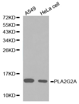
Figure 1. Western blot analysis of PLA2G2A using anti-PLA2G2A antibody (M02259-1). Electrophoresis was performed on a 5-20% SDS-PAGE gel at 70V (Stacking gel) / 90V (Resolving gel) for 2-3 hours. The sample well of each lane was loaded with 30 ug of sample under reducing conditions. Lane 1: human A549 whole cell lysates, Lane 2: human HepG2 whole cell lysates, Lane 3: human RT4 whole cell lysates, Lane 4: human Hacat whole cell lysates, Lane 5: rat small intestine tissue lysates, Lane 6: rat C6 whole cell lysates, Lane 7: mouse small intestine tissue lysates, Lane 8: mouse EL-4 whole cell lysates. After electrophoresis, proteins were transferred to a nitrocellulose membrane at 150 mA for 50-90 minutes. Blocked the membrane with 5% non-fat milk/TBS for 1.5 hour at RT. The membrane was incubated with rabbit anti-PLA2G2A antigen affinity purified monoclonal antibody (Catalog # M02259-1) at 1:500 overnight at 4°C, then washed with TBS-0.1%Tween 3 times with 5 minutes each and probed with a goat anti-rabbit IgG-HRP secondary antibody at a dilution of 1:500 for 1.5 hour at RT. The signal is developed using an Enhanced Chemiluminescent detection (ECL) kit (Catalog # EK1002) with Tanon 5200 system. A specific band was detected for PLA2G2A at approximately 16 kDa. The expected band size for PLA2G2A is at 16 kDa.
Figure 1. Western blot analysis of PLA2G2A using anti-PLA2G2A antibody (M02259-1). Electrophoresis was performed on a 5-20% SDS-PAGE gel at 70V (Stacking gel) / 90V (Resolving gel) for 2-3 hours. The sample well of each lane was loaded with 30 ug of sample under reducing conditions. Lane 1: human A549 whole cell lysates, Lane 2: human HepG2 whole cell lysates, Lane 3: human RT4 whole cell lysates, Lane 4: human Hacat whole cell lysates, Lane 5: rat small intestine tissue lysates, Lane 6: rat C6 whole cell lysates, Lane 7: mouse small intestine tissue lysates, Lane 8: mouse EL-4 whole cell lysates. After electrophoresis, proteins were transferred to a nitrocellulose membrane at 150 mA for 50-90 minutes. Blocked the membrane with 5% non-fat milk/TBS for 1.5 hour at RT. The membrane was incubated with rabbit anti-PLA2G2A antigen affinity purified monoclonal antibody (Catalog # M02259-1) at 1:500 overnight at 4°C, then washed with TBS-0.1%Tween 3 times with 5 minutes each and probed with a goat anti-rabbit IgG-HRP secondary antibody at a dilution of 1:500 for 1.5 hour at RT. The signal is developed using an Enhanced Chemiluminescent detection (ECL) kit (Catalog # EK1002) with Tanon 5200 system. A specific band was detected for PLA2G2A at approximately 16 kDa. The expected band size for PLA2G2A is at 16 kDa.
Anti-PLA2G2A Rabbit Monoclonal Antibody
M02259-1
ApplicationsWestern Blot
Product group Antibodies
ReactivityHuman, Mouse, Rat
TargetPLA2G2A
Overview
- SupplierBoster Bio
- Product NameAnti-PLA2G2A Rabbit Monoclonal Antibody
- Delivery Days Customer9
- ApplicationsWestern Blot
- CertificationResearch Use Only
- ClonalityMonoclonal
- Clone ID25P80
- Gene ID5320
- Target namePLA2G2A
- Target descriptionphospholipase A2 group IIA
- Target synonymsMOM1, PLA2, PLA2B, PLA2L, PLA2S, PLAS1, sPLA2, phospholipase A2, membrane associated, GIIC sPLA2, NPS-PLA2, group IIA phospholipase A2, non-pancreatic secretory phospholipase A2, phosphatidylcholine 2-acylhydrolase 2A, phospholipase A2, group IIA (platelets, synovial fluid)
- HostRabbit
- IsotypeIgG
- Protein IDP14555
- Protein NamePhospholipase A2, membrane associated
- Scientific DescriptionBoster Bio Anti-PLA2G2A Rabbit Monoclonal Antibody catalog # M02259-1. Tested in WB application. This antibody reacts with Human, Mouse, Rat.
- ReactivityHuman, Mouse, Rat
- Storage Instruction-20°C
- UNSPSC12352203
Related products
Product group Antibodies

PLA2G2A / SPLA2 AntibodyLS-C830881
ApplicationsELISA, ImmunoHistoChemistry
ReactivityHuman
TargetPLA2G2A
- SizePrice
Product group Antibodies
PLA2G2A AntibodyCSB-PA11209A0RB
ApplicationsELISA, ImmunoHistoChemistry
ReactivityHuman
TargetPLA2G2A
- SizePrice
Product group Antibodies

Goat anti-PLA2G2AEB12467
ApplicationsWestern Blot, ELISA, ImmunoHistoChemistry
ReactivityHuman
TargetPLA2G2A
- SizePrice
Product group Antibodies
Pla2G2A Polyclonal AntibodyCAC10412
ApplicationsELISA, ImmunoHistoChemistry
TargetPLA2G2A
- SizePrice
Product group Antibodies
Anti-PLA2G2A Antibody144-01234
ApplicationsWestern Blot
ReactivityHuman, Mouse
TargetPLA2G2A
- SizePrice
Product group Antibodies
PLA2G2A Recombinant AntibodyBSM-62378R
ApplicationsWestern Blot
ReactivityHuman, Mouse, Rat
TargetPLA2G2A
- SizePrice