


Immunohistochemical staining of human stomach, upper shows strong cytoplasmic positivity in parietal cells.
Immunohistochemical staining of human stomach, upper shows strong cytoplasmic positivity in parietal cells.
Anti-PLAC9 Antibody
HPA043469
ApplicationsImmunoHistoChemistry
Product group Antibodies
ReactivityHuman
TargetPLAC9
Overview
- SupplierAtlas Antibodies
- Product NameAnti-PLAC9 Antibody
- Delivery Days Customer4
- ApplicationsImmunoHistoChemistry
- CertificationResearch Use Only
- ClonalityPolyclonal
- ConjugateUnconjugated
- Gene ID219348
- Target namePLAC9
- Target descriptionplacenta associated 9
- Target synonymsplacenta-specific protein 9, placenta specific 9
- HostRabbit
- IsotypeIgG
- Protein IDQ5JTB6
- Protein NamePlacenta-specific protein 9
- Scientific DescriptionRecombinant Protein Epitope Signature Tag (PrEST) antigen sequence
- ReactivityHuman
- Storage Instruction-20°C,2°C to 8°C
- UNSPSC41116161
Datasheet
MSDS
Related products
Product group Antibodies

PLAC9 Antibody (Biotin)LS-C395457
ApplicationsELISA
ReactivityHuman
TargetPLAC9
- SizePrice
Product group Antibodies
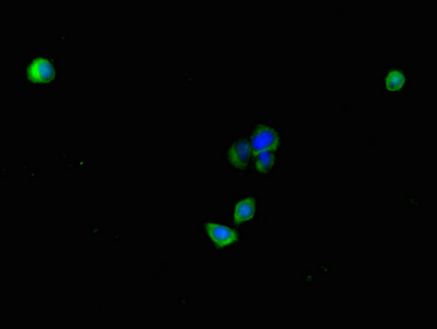
PLAC9 AntibodyCSB-PA689337LA01HU
ApplicationsImmunoFluorescence, ELISA
ReactivityHuman
TargetPLAC9
- SizePrice

Product group Antibodies
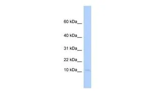
Anti-PLAC9 Antibody Picoband(r)A17853-1-CARRIER-FREE
ApplicationsWestern Blot, ELISA
ReactivityHuman
TargetPLAC9
- SizePrice
Product group Antibodies
PLAC9 antibody, InternalGTX46275
ApplicationsWestern Blot
ReactivityHuman
TargetPLAC9
- SizePrice
Product group Antibodies
PLAC9 AntibodyPACO36186
ApplicationsImmunoFluorescence, ELISA
ReactivityHuman
TargetPLAC9
- SizePrice