


Immunohistochemical staining of human lung shows strong cytoplasmic positivity in macrophages.
Immunohistochemical staining of human lung shows strong cytoplasmic positivity in macrophages.
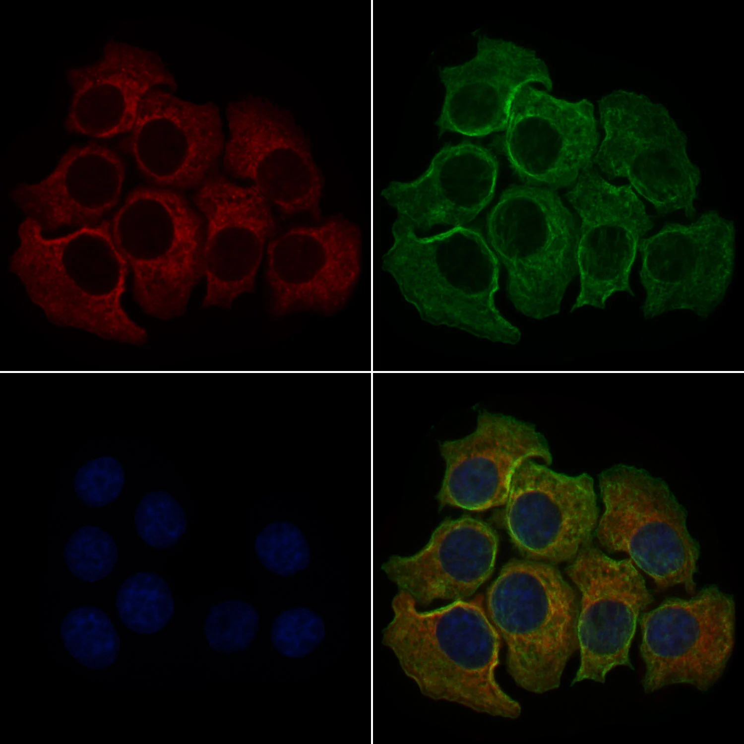
Anti-PLCE1 Antibody
HPA015597
ApplicationsImmunoHistoChemistry
Product group Antibodies
ReactivityHuman
TargetPLCE1
Overview
- SupplierAtlas Antibodies
- Product NameAnti-PLCE1 Antibody
- Delivery Days Customer4
- ApplicationsImmunoHistoChemistry
- CertificationResearch Use Only
- ClonalityPolyclonal
- ConjugateUnconjugated
- Gene ID51196
- Target namePLCE1
- Target descriptionphospholipase C epsilon 1
- Target synonymsNPHS3, PLCE, PPLC, 1-phosphatidylinositol 4,5-bisphosphate phosphodiesterase epsilon-1, PLC-epsilon-1, pancreas-enriched phospholipase C, phosphoinositide phospholipase C, phosphoinositide phospholipase C-epsilon-1, phosphoinositide-specific phospholipase C epsilon-1
- HostRabbit
- IsotypeIgG
- Protein IDQ9P212
- Protein Name1-phosphatidylinositol 4,5-bisphosphate phosphodiesterase epsilon-1
- Scientific DescriptionRecombinant Protein Epitope Signature Tag (PrEST) antigen sequence
- ReactivityHuman
- Storage Instruction-20°C,2°C to 8°C
- UNSPSC41116161
Datasheet
MSDS
Related products
Product group Antibodies
PLCE1 antibodyGTX02580
ApplicationsImmunoFluorescence, Western Blot, ImmunoCytoChemistry, ImmunoHistoChemistry, ImmunoHistoChemistry Paraffin
ReactivityHuman, Monkey, Mouse, Rat
TargetPLCE1
- SizePrice
Product group Antibodies

PLCE1 AntibodyLS-C663427
ApplicationsImmunoPrecipitation, Western Blot, ImmunoCytoChemistry
ReactivityMouse
TargetPLCE1
- SizePrice

Product group Antibodies
Anti-PLCE1 Antibody Picoband(r)A02360-1-CARRIER-FREE
ApplicationsWestern Blot, ELISA
ReactivityHuman
TargetPLCE1
- SizePrice