Bio-Connect
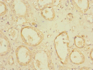
Immunohistochemical staining of human duodenum shows moderate to strong nuclear positivity in glandular cells.

Anti-POGK Antibody

HPA031630
Atlas Antibodies
ApplicationsImmunoCytoChemistry, ImmunoHistoChemistry
Product group Antibodies
ReactivityHuman
TargetPOGK
Sign in to order and to see your custom pricing.
Large volume orders?
Order with a bulk request

Overview

  • Supplier
    Atlas Antibodies
  • Product Name
    Anti-POGK
  • Delivery Days Customer
    4
  • Applications
    ImmunoCytoChemistry, ImmunoHistoChemistry
  • Certification
    Research Use Only
  • Clonality
    Polyclonal
  • Conjugate
    Unconjugated
  • Gene ID57645
  • Target name
    POGK
  • Target description
    pogo transposable element derived with KRAB domain
  • Target synonyms
    BASS2; KRAB box domain containing 2; KRBOX2; LST003; pogo transposable element with KRAB domain
  • Host
    Rabbit
  • Isotype
    IgG
  • Protein IDQ9P215
  • Protein Name
    Pogo transposable element with KRAB domain
  • Scientific Description
    Recombinant Protein Epitope Signature Tag (PrEST) antigen sequence
  • Reactivity
    Human
  • Storage Instruction
    -20°C,2°C to 8°C
  • UNSPSC
    41116161

Related products

Product group Antibodies
Bethyl Laboratories
ApplicationsImmunoPrecipitation, Western Blot
TargetPOGK
  • SizePrice
Immunohistochemistry of paraffin-embedded human kidney tissue using CSB-PA018298LA01HU at dilution of 1:100
Product group Antibodies
Cusabio
POGK AntibodyCSB-PA018298LA01HU
ApplicationsImmunoFluorescence, ELISA, ImmunoHistoChemistry
ReactivityHuman
TargetPOGK
  • SizePrice
Product group Antibodies
LifeSpan BioSciences
ApplicationsELISA
TargetPOGK
  • SizePrice
Product group Antibodies
Biosynth
  • SizePrice
Atlas Antibodies
Protein IDQ9P215
  • SizePrice
Figure 1. Western blot analysis of POGK using anti-POGK antibody (A15311-1). Electrophoresis was performed on a 5-20% SDS-PAGE gel at 70V (Stacking gel) / 90V (Resolving gel) for 2-3 hours. The sample well of each lane was loaded with 30 ug of sample under reducing conditions. Lane 1: human A549 whole cell lysates, Lane 2: human MCF-7 whole cell lysates, Lane 3: human RT4 whole cell lysates, Lane 4: human SH-SY5Y whole cell lysates, Lane 5: rat brain tissue lysates, Lane 6: rat testis tissue lysates, Lane 7: mouse brain tissue lysates, Lane 8: mouse testis tissue lysates. After electrophoresis, proteins were transferred to a nitrocellulose membrane at 150 mA for 50-90 minutes. Blocked the membrane with 5% non-fat milk/TBS for 1.5 hour at RT. The membrane was incubated with rabbit anti-POGK antigen affinity purified polyclonal antibody (Catalog # A15311-1) at 0.5 microg/mL overnight at 4°C, then washed with TBS-0.1%Tween 3 times with 5 minutes each and probed with a goat anti-rabbit IgG-HRP secondary antibody at a dilution of 1:5000 for 1.5 hour at RT. The signal is developed using an Enhanced Chemiluminescent detection (ECL) kit (Catalog # EK1002) with Tanon 5200 system. A specific band was detected for POGK at approximately 95 kDa. The expected band size for POGK is at 69 kDa.
Product group Antibodies
Boster Bio
Anti-POGK Antibody Picoband(r)A15311-1-CARRIER-FREE
ApplicationsFlow Cytometry, ImmunoFluorescence, Western Blot, ELISA, ImmunoCytoChemistry
TargetPOGK
  • SizePrice
Product group Antibodies
Assay Genie
POGK AntibodyPACO29042
ApplicationsImmunoFluorescence, ELISA, ImmunoHistoChemistry
TargetPOGK
  • SizePrice