


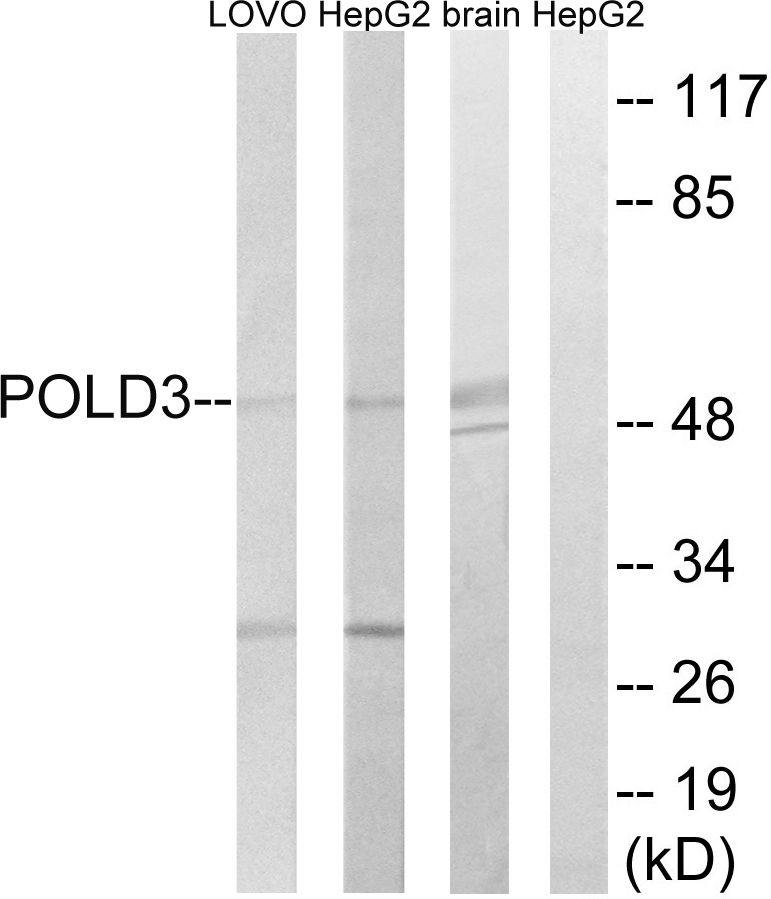
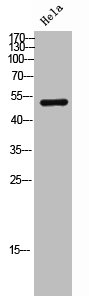
Figure 1. Western blot analysis of POLD3 using anti-POLD3 antibody (A05111-2). Electrophoresis was performed on a 5-20% SDS-PAGE gel at 70V (Stacking gel) / 90V (Resolving gel) for 2-3 hours. The sample well of each lane was loaded with 30 ug of sample under reducing conditions. Lane 1: human MCF-7 whole cell lysates, Lane 2: human Hela whole cell lysates, Lane 3: rat testis tissue lysates, Lane 4: mouse testis tissue lysates. After electrophoresis, proteins were transferred to a nitrocellulose membrane at 150 mA for 50-90 minutes. Blocked the membrane with 5% non-fat milk/TBS for 1.5 hour at RT. The membrane was incubated with rabbit anti-POLD3 antigen affinity purified polyclonal antibody (Catalog # A05111-2) at 0.5 microg/mL overnight at 4°C, then washed with TBS-0.1%Tween 3 times with 5 minutes each and probed with a goat anti-rabbit IgG-HRP secondary antibody at a dilution of 1:5000 for 1.5 hour at RT. The signal is developed using an Enhanced Chemiluminescent detection (ECL) kit (Catalog # EK1002) with Tanon 5200 system. A specific band was detected for POLD3 at approximately 70 kDa. The expected band size for POLD3 is at 51 kDa.
Figure 1. Western blot analysis of POLD3 using anti-POLD3 antibody (A05111-2). Electrophoresis was performed on a 5-20% SDS-PAGE gel at 70V (Stacking gel) / 90V (Resolving gel) for 2-3 hours. The sample well of each lane was loaded with 30 ug of sample under reducing conditions. Lane 1: human MCF-7 whole cell lysates, Lane 2: human Hela whole cell lysates, Lane 3: rat testis tissue lysates, Lane 4: mouse testis tissue lysates. After electrophoresis, proteins were transferred to a nitrocellulose membrane at 150 mA for 50-90 minutes. Blocked the membrane with 5% non-fat milk/TBS for 1.5 hour at RT. The membrane was incubated with rabbit anti-POLD3 antigen affinity purified polyclonal antibody (Catalog # A05111-2) at 0.5 microg/mL overnight at 4°C, then washed with TBS-0.1%Tween 3 times with 5 minutes each and probed with a goat anti-rabbit IgG-HRP secondary antibody at a dilution of 1:5000 for 1.5 hour at RT. The signal is developed using an Enhanced Chemiluminescent detection (ECL) kit (Catalog # EK1002) with Tanon 5200 system. A specific band was detected for POLD3 at approximately 70 kDa. The expected band size for POLD3 is at 51 kDa.
Anti-POLD3 Antibody Picoband(r)
A05111-2-CARRIER-FREE
ApplicationsWestern Blot, ELISA
Product group Antibodies
ReactivityHuman, Mouse, Rat
TargetPOLD3
Overview
- SupplierBoster Bio
- Product NameAnti-POLD3 Antibody Picoband(r)
- Delivery Days Customer9
- ApplicationsWestern Blot, ELISA
- CertificationResearch Use Only
- ClonalityPolyclonal
- Concentration500 ug/ml
- Gene ID10714
- Target namePOLD3
- Target descriptionDNA polymerase delta 3, accessory subunit
- Target synonymsIMD122, P66, P68, PPP1R128, DNA polymerase delta subunit 3, DNA polymerase delta subunit C, DNA polymerase delta subunit p66, DNA polymerase delta subunit p68, Pol delta C subunit (p66), polymerase (DNA) delta 3, accessory subunit, polymerase (DNA-directed), delta 3, accessory subunit, protein phosphatase 1, regulatory subunit 128
- HostRabbit
- Protein IDQ15054
- Protein NameDNA polymerase delta subunit 3
- Scientific DescriptionBoster Bio Anti-POLD3 Antibody Picoband® catalog # A05111-2. Tested in WB, ELISA applications. This antibody reacts with Human, Mouse, Rat. The brand Picoband indicates this is a premium antibody that guarantees superior quality, high affinity, and strong signals with minimal background in Western blot applications. Only our best-performing antibodies are designated as Picoband, ensuring unmatched performance.
- ReactivityHuman, Mouse, Rat
- Storage Instruction-20°C,2°C to 8°C
- UNSPSC12352203
Related products
Product group Antibodies
Anti-POLD3 AntibodyA97341
ApplicationsWestern Blot, ELISA
ReactivityHuman, Mouse, Rat
- SizePrice
Product group Antibodies
Anti-POLD3 Antibody144-07243
ApplicationsImmunoFluorescence, Western Blot, ImmunoHistoChemistry
ReactivityHuman, Mouse
TargetPOLD3
- SizePrice
Product group Antibodies
POLD3 AntibodyCSB-PA006684
ApplicationsWestern Blot, ELISA
ReactivityHuman, Mouse, Rat
TargetPOLD3
- SizePrice
Product group Antibodies
Pold3 Polyclonal AntibodyCAC11251
ApplicationsImmunoFluorescence, Western Blot, ELISA, ImmunoHistoChemistry
ReactivityMouse
TargetPOLD3
- SizePrice
Product group Antibodies

POLD3 AntibodyLS-C346302
ApplicationsImmunoFluorescence, Western Blot, ImmunoHistoChemistry
ReactivityHuman
TargetPOLD3
- SizePrice

Product group Antibodies
Anti-POLD3 AntibodyHPA058846
ApplicationsImmunoCytoChemistry, ImmunoHistoChemistry
ReactivityHuman
TargetPOLD3
- SizePrice
Product group Antibodies
POLD3 antibodyGTX32795
ApplicationsImmunoFluorescence, Western Blot, ImmunoCytoChemistry, ImmunoHistoChemistry, ImmunoHistoChemistry Paraffin
ReactivityHuman, Mouse
TargetPOLD3
- SizePrice
Product group Antibodies
Anti-POLD3 AntibodyCAB7243
ApplicationsImmunoFluorescence, Western Blot, ELISA, ImmunoCytoChemistry, ImmunoHistoChemistry, ImmunoHistoChemistry Paraffin
ReactivityHuman
TargetPOLD3
- SizePrice