

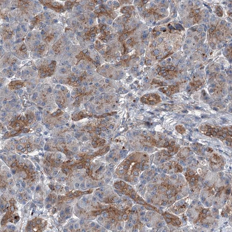
Immunohistochemical staining of human pancreas shows moderate cytoplasmic positivity in exocrine glandular cells and islets of Langerhans.
 and POLR1D over-expression lysate (Co-expressed with a C-terminal myc-DDK tag (~3.1 kDa) in mammalian HEK293T cells, LY407348).")
Immunohistochemical staining of human pancreas shows moderate cytoplasmic positivity in exocrine glandular cells and islets of Langerhans.
Anti-POLR1D Antibody
HPA039337
ApplicationsWestern Blot, ImmunoCytoChemistry, ImmunoHistoChemistry
Product group Antibodies
ReactivityHuman
TargetPOLR1D
Overview
- SupplierAtlas Antibodies
- Product NameAnti-POLR1D Antibody
- Delivery Days Customer4
- ApplicationsWestern Blot, ImmunoCytoChemistry, ImmunoHistoChemistry
- CertificationResearch Use Only
- ClonalityPolyclonal
- ConjugateUnconjugated
- Gene ID51082
- Target namePOLR1D
- Target descriptionRNA polymerase I and III subunit D
- Target synonymsAC19, POLR1C, RPA16, RPA9, RPAC2, RPC16, RPO1-3, TCS2, DNA-directed RNA polymerases I and III subunit RPAC2, Protein POLR1D, DNA-directed RNA polymerase I subunit D, RNA polymerase I subunit D, RNA polymerases I and III subunit AC2, polymerase (RNA) I polypeptide D, 16kDa, polymerase (RNA) I subunit D
- HostRabbit
- IsotypeIgG
- Protein IDQ9Y2S0
- Protein NameDNA-directed RNA polymerases I and III subunit RPAC2
- Scientific DescriptionRecombinant Protein Epitope Signature Tag (PrEST) antigen sequence
- ReactivityHuman
- Storage Instruction-20°C,2°C to 8°C
- UNSPSC41116161
Datasheet
MSDS
Related products

Product group Antibodies
Anti-POLR1D Antibody Picoband(r)A07808-1-CARRIER-FREE
ApplicationsWestern Blot, ELISA
TargetPOLR1D
- SizePrice
Product group Antibodies
Anti-POLR1D Antibody144-08021
ApplicationsImmunoFluorescence, Western Blot
TargetPOLR1D
- SizePrice
Product group Antibodies
POLR1D antibody [N1C3]GTX115993
ApplicationsWestern Blot, ImmunoHistoChemistry, ImmunoHistoChemistry Paraffin
TargetPOLR1D
- SizePrice
Product group Antibodies

POLR1D AntibodyLS-C832164
ApplicationsELISA, ImmunoHistoChemistry
TargetPOLR1D
- SizePrice
Product group Antibodies
POLR1D AntibodyCSB-PA897095ESR1HU
ApplicationsELISA, ImmunoHistoChemistry
ReactivityHuman
TargetPOLR1D
- SizePrice