

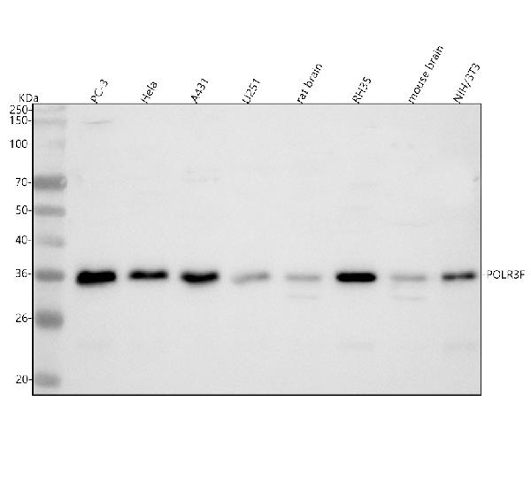
Figure 1. Western blot analysis of POLR3F using anti-POLR3F antibody (A12720-1). Electrophoresis was performed on a 5-20% SDS-PAGE gel at 70V (Stacking gel) / 90V (Resolving gel) for 2-3 hours. The sample well of each lane was loaded with 30 ug of sample under reducing conditions. Lane 1: human PC-3 whole cell lysates, Lane 2: human Hela whole cell lysates, Lane 3: human A431 whole cell lysates, Lane 4: human U251 whole cell lysates, Lane 5: rat brain tissue lysates, Lane 6: rat RH35 whole cell lysates, Lane 7: mouse brain tissue lysates, Lane 8: mouse NIH/3T3 whole cell lysates. After electrophoresis, proteins were transferred to a nitrocellulose membrane at 150 mA for 50-90 minutes. Blocked the membrane with 5% non-fat milk/TBS for 1.5 hour at RT. The membrane was incubated with rabbit anti-POLR3F antigen affinity purified polyclonal antibody (Catalog # A12720-1) at 0.5 microg/mL overnight at 4°C, then washed with TBS-0.1%Tween 3 times with 5 minutes each and probed with a goat anti-rabbit IgG-HRP secondary antibody at a dilution of 1:5000 for 1.5 hour at RT. The signal is developed using an Enhanced Chemiluminescent detection (ECL) kit (Catalog # EK1002) with Tanon 5200 system. A specific band was detected for POLR3F at approximately 36 kDa. The expected band size for POLR3F is at 36 kDa.
 and anti-Beta Tubulin antibody (M01857-3). POLR3F was detected in immunocytochemical section of MCF-7 cell. Enzyme antigen retrieval was performed using IHC enzyme antigen retrieval reagent (AR0022) for 15 mins. The cells were blocked with 10% goat serum. And then incubated with 5 microg/mL rabbit anti-POLR3F Antibody (A12720-1) and mouse anti-Beta Tubulin antibody (M01857-3) overnight at 4°C. DyLight®488 Conjugated Goat Anti-Rabbit IgG (BA1127)(B) and DyLight®550 Conjugated Goat Anti-Mouse IgG (BA1133)(D) were used as secondary antibody at 1:500 dilution and incubated for 30 minutes at 37°C. The section was counterstained with DAPI(C). Visualize using a fluorescence microscope and filter sets appropriate for the label used.")
. Overlay histogram showing A431 cells stained with A12720-1 (Blue line). To facilitate intracellular staining, cells were fixed with 4% paraformaldehyde and permeabilized with permeabilization buffer. The cells were blocked with 10% normal goat serum. And then incubated with rabbit anti-POLR3F Antibody (A12720-1, 1 microg/1x106 cells) for 30 min at 20°C. DyLight®488 conjugated goat anti-rabbit IgG (BA1127, 5-10 microg/1x106 cells) was used as secondary antibody for 30 minutes at 20°C. Isotype control antibody (Green line) was rabbit IgG (1 microg/1x106) used under the same conditions. Unlabelled sample (Red line) was also used as a control.")
Figure 1. Western blot analysis of POLR3F using anti-POLR3F antibody (A12720-1). Electrophoresis was performed on a 5-20% SDS-PAGE gel at 70V (Stacking gel) / 90V (Resolving gel) for 2-3 hours. The sample well of each lane was loaded with 30 ug of sample under reducing conditions. Lane 1: human PC-3 whole cell lysates, Lane 2: human Hela whole cell lysates, Lane 3: human A431 whole cell lysates, Lane 4: human U251 whole cell lysates, Lane 5: rat brain tissue lysates, Lane 6: rat RH35 whole cell lysates, Lane 7: mouse brain tissue lysates, Lane 8: mouse NIH/3T3 whole cell lysates. After electrophoresis, proteins were transferred to a nitrocellulose membrane at 150 mA for 50-90 minutes. Blocked the membrane with 5% non-fat milk/TBS for 1.5 hour at RT. The membrane was incubated with rabbit anti-POLR3F antigen affinity purified polyclonal antibody (Catalog # A12720-1) at 0.5 microg/mL overnight at 4°C, then washed with TBS-0.1%Tween 3 times with 5 minutes each and probed with a goat anti-rabbit IgG-HRP secondary antibody at a dilution of 1:5000 for 1.5 hour at RT. The signal is developed using an Enhanced Chemiluminescent detection (ECL) kit (Catalog # EK1002) with Tanon 5200 system. A specific band was detected for POLR3F at approximately 36 kDa. The expected band size for POLR3F is at 36 kDa.
Anti-POLR3F Antibody Picoband(r)
A12720-1-CARRIER-FREE
ApplicationsFlow Cytometry, ImmunoFluorescence, Western Blot, ELISA, ImmunoCytoChemistry
Product group Antibodies
ReactivityHuman, Mouse, Rat
TargetPOLR3F
Overview
- SupplierBoster Bio
- Product NameAnti-POLR3F Antibody Picoband(r)
- Delivery Days Customer9
- ApplicationsFlow Cytometry, ImmunoFluorescence, Western Blot, ELISA, ImmunoCytoChemistry
- CertificationResearch Use Only
- ClonalityPolyclonal
- Concentration500 ug/ml
- Gene ID10621
- Target namePOLR3F
- Target descriptionRNA polymerase III subunit F
- Target synonymsC34, IMD101, RPC39, RPC6, DNA-directed RNA polymerase III subunit RPC6, RNA polymerase III C39 subunit, RNA polymerase III subunit C6, polymerase (RNA) III (DNA directed) polypeptide F, 39 kDa, polymerase (RNA) III subunit F
- HostRabbit
- Protein IDQ9H1D9
- Protein NameDNA-directed RNA polymerase III subunit RPC6
- Scientific DescriptionBoster Bio Anti-POLR3F Antibody Picoband® catalog # A12720-1. Tested in WB, ICC/IF, Flow Cytometry, ELISA applications. This antibody reacts with Human, Mouse, Rat. The brand Picoband indicates this is a premium antibody that guarantees superior quality, high affinity, and strong signals with minimal background in Western blot applications. Only our best-performing antibodies are designated as Picoband, ensuring unmatched performance.
- ReactivityHuman, Mouse, Rat
- Storage Instruction-20°C,2°C to 8°C
- UNSPSC12352203
Related products
Product group Antibodies
Anti-POLR3F AntibodyA28339
ApplicationsWestern Blot
ReactivityHuman, Mouse, Rat
- SizePrice
Product group Antibodies

POLR3F AntibodyLS-C830734
ApplicationsWestern Blot, ELISA, ImmunoHistoChemistry
ReactivityHuman, Mouse, Rat
TargetPOLR3F
- SizePrice

Product group Antibodies
Anti-POLR3F AntibodyHPA049441
ApplicationsWestern Blot, ImmunoHistoChemistry
ReactivityHuman
TargetPOLR3F
- SizePrice
Product group Antibodies
POLR3F AntibodyCSB-PA884440LA01HU
ApplicationsWestern Blot, ELISA
ReactivityHuman
TargetPOLR3F
- SizePrice
Product group Antibodies
POLR3F Polyclonal AntibodyCAC14467
ApplicationsWestern Blot, ELISA
TargetPOLR3F
- SizePrice
Product group Antibodies
POLR3F antibodyGTX105818
ApplicationsWestern Blot
ReactivityHuman
TargetPOLR3F
- SizePrice
Product group Antibodies
Anti-POLR3F Antibody144-12224
ApplicationsWestern Blot
ReactivityHuman, Mouse, Rat
TargetPOLR3F
- SizePrice