


Immunohistochemical staining of human kidney shows moderate cytoplasmic positivity in cells in tubules.
Immunohistochemical staining of human kidney shows moderate cytoplasmic positivity in cells in tubules.
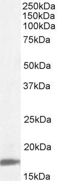
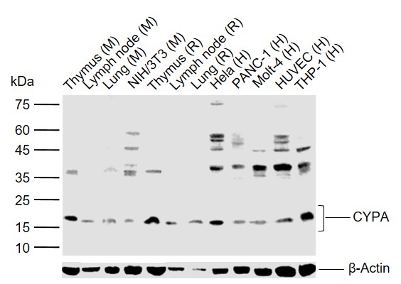
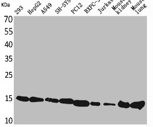
Anti-PPIA Antibody
HPA058345
ApplicationsImmunoHistoChemistry
Product group Antibodies
ReactivityHuman
TargetPPIA
Overview
- SupplierAtlas Antibodies
- Product NameAnti-PPIA Antibody
- Delivery Days Customer4
- ApplicationsImmunoHistoChemistry
- CertificationResearch Use Only
- ClonalityPolyclonal
- ConjugateUnconjugated
- Gene ID5478
- Target namePPIA
- Target descriptionpeptidylprolyl isomerase A
- Target synonymsCYPA, CYPH, HEL-S-69p, peptidyl-prolyl cis-trans isomerase A, PPIase A, T cell cyclophilin, cyclosporin A-binding protein, epididymis secretory sperm binding protein Li 69p, peptidylprolyl isomerase A (cyclophilin A), rotamase A
- HostRabbit
- IsotypeIgG
- Protein IDP62937
- Protein NamePeptidyl-prolyl cis-trans isomerase A
- Scientific DescriptionRecombinant Protein Epitope Signature Tag (PrEST) antigen sequence
- ReactivityHuman
- Storage Instruction-20°C,2°C to 8°C
- UNSPSC41116161
Datasheet
MSDS
Related products
Product group Antibodies
ApplicationsWestern Blot, ELISA
ReactivityHuman
- SizePrice

Product group Antibodies
Anti-Cyclophilin A/PPIA Antibody Picoband(r)A01308-CARRIER-FREE
ApplicationsFlow Cytometry, ImmunoFluorescence, Western Blot, ImmunoCytoChemistry, ImmunoHistoChemistry, ImmunoHistoChemistry Frozen
ReactivityHuman, Mouse, Rat
TargetPPIA
- SizePrice
Product group Antibodies
Anti-PPIA Antibody144-00993
ApplicationsWestern Blot
ReactivityHuman, Mouse, Rat
TargetPPIA
- SizePrice
Product group Antibodies
CYPA Polyclonal AntibodyBS-5912R
ApplicationsFlow Cytometry, ImmunoFluorescence, Western Blot, ELISA, ImmunoCytoChemistry, ImmunoHistoChemistry, ImmunoHistoChemistry Frozen, ImmunoHistoChemistry Paraffin
ReactivityBovine, Canine, Human, Mouse, Porcine, Rabbit, Rat
TargetPPIA
- SizePrice
Product group Antibodies

ApplicationsWestern Blot, ELISA
ReactivityBovine, Canine, Human, Mouse, Porcine, Rat
TargetPPIA
- SizePrice
Product group Antibodies
PPIA Polyclonal AntibodyCAC14102
ApplicationsWestern Blot, ELISA
ReactivityMouse
TargetPPIA
- SizePrice
Product group Antibodies
PPIA AntibodyCSB-PA005117
ApplicationsWestern Blot, ELISA
ReactivityHuman, Mouse, Rat
TargetPPIA
- SizePrice
Product group Antibodies

PPIA / Cyclophilin A AntibodyLS-C405253
ApplicationsWestern Blot, ELISA, ImmunoHistoChemistry
ReactivityHuman, Mouse, Rat
TargetPPIA
- SizePrice