

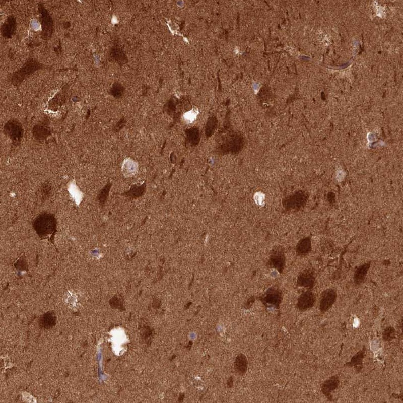
Immunohistochemical staining of human hippocampus shows strong cytoplasmic positivity in neuronal cells.

Immunohistochemical staining of human hippocampus shows strong cytoplasmic positivity in neuronal cells.
Anti-PPP3R1 Antibody
HPA047157
ApplicationsImmunoCytoChemistry, ImmunoHistoChemistry
Product group Antibodies
ReactivityHuman
TargetPPP3R1
Overview
- SupplierAtlas Antibodies
- Product NameAnti-PPP3R1 Antibody
- Delivery Days Customer4
- ApplicationsImmunoCytoChemistry, ImmunoHistoChemistry
- CertificationResearch Use Only
- ClonalityPolyclonal
- ConjugateUnconjugated
- Gene ID5534
- Target namePPP3R1
- Target descriptionprotein phosphatase 3 regulatory subunit B, alpha
- Target synonymsCALNB1, CNB, CNB1, calcineurin subunit B type 1, calcineurin B, type I (19kDa), protein phosphatase 2B regulatory subunit 1, protein phosphatase 2B regulatory subunit B alpha
- HostRabbit
- IsotypeIgG
- Protein IDP63098
- Protein NameCalcineurin subunit B type 1
- Scientific DescriptionRecombinant Protein Epitope Signature Tag (PrEST) antigen sequence
- ReactivityHuman
- Storage Instruction-20°C,2°C to 8°C
- UNSPSC41116161
Datasheet
MSDS
Related products

Product group Antibodies
Anti-PPP3R1 Antibody Picoband(r)A06469-1-CARRIER-FREE
ApplicationsFlow Cytometry, Western Blot, ELISA, ImmunoHistoChemistry
ReactivityHuman, Mouse, Rat
TargetPPP3R1
- SizePrice
Product group Antibodies
Anti-PPP3R1 Antibody144-00954
ApplicationsWestern Blot, ImmunoHistoChemistry
ReactivityHuman, Mouse, Rat
TargetPPP3R1
- SizePrice
Product group Antibodies

PPP3R1 / Calcineurin B AntibodyLS-C831934
ApplicationsELISA, ImmunoHistoChemistry
ReactivityHuman, Mouse, Rat
TargetPPP3R1
- SizePrice
Product group Antibodies
ApplicationsImmunoFluorescence, ELISA, ImmunoCytoChemistry, ImmunoHistoChemistry, ImmunoHistoChemistry Frozen, ImmunoHistoChemistry Paraffin
ReactivityBovine, Canine, Drosophila, Equine, Guinea Pig, Human, Mouse, Porcine, Rabbit, Rat, Sheep, Zebra Fish
TargetPPP3R1
- SizePrice
Product group Antibodies
PPP3R1 Polyclonal AntibodyCAC13882
ApplicationsWestern Blot, ELISA
TargetPPP3R1
- SizePrice
Product group Antibodies
PPP3R1 AntibodyCSB-PA018575LA01HU
ApplicationsWestern Blot, ELISA
ReactivityHuman
TargetPPP3R1
- SizePrice
Product group Antibodies
Calcineurin B antibodyGTX109127
ApplicationsWestern Blot, ImmunoHistoChemistry, ImmunoHistoChemistry Paraffin
ReactivityHuman, Mouse, Rat, Zebra Fish
TargetPPP3R1
- SizePrice
Product group Antibodies
Anti-PPP3R1 AntibodyCAB0954
ApplicationsImmunoFluorescence, Western Blot, ELISA, ImmunoCytoChemistry
ReactivityHuman
TargetPPP3R1
- SizePrice