


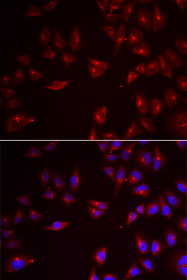
Immunofluorescence staining of U-2 OS cells using the Anti-PRAME monoclonal antibody, showing specific staining in the nucleus and cytosol in green. Microtubule- and nuclear probes are visualized in red and blue, respectively (where available).

Immunofluorescence staining of U-2 OS cells using the Anti-PRAME monoclonal antibody, showing specific staining in the nucleus and cytosol in green. Microtubule- and nuclear probes are visualized in red and blue, respectively (where available).
Anti-PRAME Antibody
AMAB91329
ApplicationsWestern Blot, ImmunoCytoChemistry
Product group Antibodies
ReactivityHuman
TargetPRAME
Overview
- SupplierAtlas Antibodies
- Product NameAnti-PRAME Antibody
- Delivery Days Customer4
- ApplicationsWestern Blot, ImmunoCytoChemistry
- CertificationResearch Use Only
- ClonalityMonoclonal
- Clone IDCL5146
- ConjugateUnconjugated
- Gene ID23532
- Target namePRAME
- Target descriptionPRAME nuclear receptor transcriptional regulator
- Target synonymsCT130, MAPE, OIP-4, OIP4, melanoma antigen preferentially expressed in tumors, Opa-interacting protein OIP4, cancer/testis antigen 130, opa-interacting protein 4, preferentially expressed antigen in melanoma, preferentially expressed antigen of melanoma
- HostMouse
- IsotypeIgG2a
- Protein IDP78395
- Protein NameMelanoma antigen preferentially expressed in tumors
- Scientific DescriptionRecombinant Protein Epitope Signature Tag (PrEST) antigen sequence
- ReactivityHuman
- Storage Instruction-20°C,2°C to 8°C
- UNSPSC41116161
Datasheet
MSDS
Related products
Product group Antibodies
Anti-PRAME AntibodyA31396
ApplicationsImmunoFluorescence, Western Blot, ImmunoHistoChemistry
ReactivityHuman
- SizePrice

Product group Antibodies
Anti-PRAME Picoband(r) AntibodyA06628-2-CARRIER-FREE
ApplicationsWestern Blot, ELISA
ReactivityHuman
TargetPRAME
- SizePrice
Product group Antibodies
Anti-PRAME Antibody144-60684
ApplicationsWestern Blot
ReactivityHuman, Mouse, Rat
TargetPRAME
- SizePrice

Product group Antibodies
Anti-PRAME AntibodyAMAB91330
ApplicationsWestern Blot, ImmunoHistoChemistry
ReactivityHuman
TargetPRAME
- SizePrice
Product group Antibodies

PRAME AntibodyLS-C831773
ApplicationsELISA, ImmunoHistoChemistry
ReactivityHuman
TargetPRAME
- SizePrice
Product group Antibodies
PRAME Polyclonal AntibodyBS-7004R
ApplicationsFlow Cytometry, ImmunoFluorescence, ELISA, ImmunoCytoChemistry, ImmunoHistoChemistry, ImmunoHistoChemistry Frozen, ImmunoHistoChemistry Paraffin
ReactivityHuman, Mouse
TargetPRAME
- SizePrice
Product group Antibodies
Prame Polyclonal AntibodyCAC08754
ApplicationsELISA, ImmunoHistoChemistry
TargetPRAME
- SizePrice
Product group Antibodies
PRAME AntibodyCSB-PA018603LA01HU
ApplicationsELISA, ImmunoHistoChemistry
ReactivityHuman
TargetPRAME
- SizePrice