


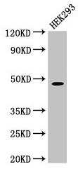
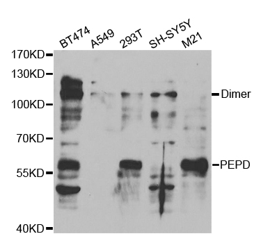
Western blot analysis of PRD expression in HepG2 cell lysate.
Western blot analysis of PRD expression in HepG2 cell lysate.
Anti-PRD Rabbit Monoclonal Antibody
M03417-2
ApplicationsWestern Blot
Product group Antibodies
ReactivityHuman
TargetPEPD
Overview
- SupplierBoster Bio
- Product NameAnti-PRD Rabbit Monoclonal Antibody
- Delivery Days Customer9
- ApplicationsWestern Blot
- CertificationResearch Use Only
- ClonalityMonoclonal
- Clone ID30P28
- Gene ID5184
- Target namePEPD
- Target descriptionpeptidase D
- Target synonymsPROLIDASE, xaa-Pro dipeptidase, X-Pro dipeptidase, aminoacyl-L-proline hydrolase, imidodipeptidase, proline dipeptidase, testicular tissue protein Li 138
- HostRabbit
- IsotypeIgG
- Protein IDP12955
- Protein NameXaa-Pro dipeptidase
- Scientific DescriptionBoster Bio Anti-PRD Rabbit Monoclonal Antibody catalog # M03417-2. Tested in WB application. This antibody reacts with Human.
- ReactivityHuman
- Storage Instruction-20°C
- UNSPSC12352203
Related products

Product group Antibodies
Anti-PEPD Antibody Picoband(r)A03417-1-CARRIER-FREE
ApplicationsFlow Cytometry, ImmunoFluorescence, Western Blot, ELISA, ImmunoCytoChemistry
ReactivityHuman, Mouse, Rat
TargetPEPD
- SizePrice
Product group Antibodies
Anti-PEPD Antibody144-05416
ApplicationsWestern Blot
ReactivityHuman, Mouse, Rat
TargetPEPD
- SizePrice
Product group Antibodies
PEPD Polyclonal AntibodyCAC14748
ApplicationsWestern Blot, ELISA, ImmunoHistoChemistry
TargetPEPD
- SizePrice
Product group Antibodies
ApplicationsFlow Cytometry, Western Blot, ImmunoCytoChemistry
ReactivityHuman
TargetPEPD
- SizePrice
Product group Antibodies
PEPD AntibodyCSB-PA017784LA01HU
ApplicationsWestern Blot, ELISA, ImmunoHistoChemistry
ReactivityHuman
TargetPEPD
- SizePrice

Product group Antibodies
Anti-PEPD AntibodyHPA072045
ApplicationsImmunoCytoChemistry
ReactivityHuman
TargetPEPD
- SizePrice
Product group Antibodies
Anti-PEPDY058828
ApplicationsWestern Blot, ImmunoHistoChemistry
ReactivityHuman
- SizePrice
Product group Antibodies
Anti-PEPD AntibodyA30778
ApplicationsWestern Blot, ImmunoHistoChemistry
ReactivityHuman, Mouse, Rat
- SizePrice