


Figure 1. Western blot analysis of PRDM14 using anti-PRDM14 antibody (A03870-2). Electrophoresis was performed on a 5-20% SDS-PAGE gel at 70V (Stacking gel) / 90V (Resolving gel) for 2-3 hours. The sample well of each lane was loaded with 30ug of sample under reducing conditions. Lane 1: rat testis tissue lysates, Lane 2: rat spleen tissue lysates, Lane 3: rat C6 whole cell lysates, Lane 4: rat PC-12 whole cell lysates, Lane 5: mouse testis tissue lysates, Lane 6: mouse spleen tissue lysates, Lane 7: mouse NIH/3T3 whole cell lysates, Lane 8: mouse Neuro-2a whole cell lysates. After Electrophoresis, proteins were transferred to a Nitrocellulose membrane at 150mA for 50-90 minutes. Blocked the membrane with 5% Non-fat Milk/ TBS for 1.5 hour at RT. The membrane was incubated with rabbit anti-PRDM14 antigen affinity purified polyclonal antibody (Catalog # A03870-2) at 0.25 microg/mL overnight at 4°C, then washed with TBS-0.1%Tween 3 times with 5 minutes each and probed with a goat anti-rabbit IgG-HRP secondary antibody at a dilution of 1:5000 for 1.5 hour at RT. The signal is developed using an Enhanced Chemiluminescent detection (ECL) kit (Catalog # EK1002) with Tanon 5200 system. A specific band was detected for PRDM14 at approximately 64KD. The expected band size for PRDM14 is at 64KD.
. Overlay histogram showing SiHa cells stained with A03870-2 (Blue line). To facilitate intracellular staining, cells were fixed with 4% paraformaldehyde and permeabilized with permeabilization buffer. The cells were blocked with 10% normal goat serum. And then incubated with rabbit anti-PRDM14 Antibody (A03870-2, 1microg/1x106 cells) for 30 min at 20°C. DyLight®488 conjugated goat anti-rabbit IgG (BA1127, 5-10microg/1x106 cells) was used as secondary antibody for 30 minutes at 20°C. Isotype control antibody (Green line) was rabbit IgG (1microg/1x106) used under the same conditions. Unlabelled sample without incubation with primary antibody and secondary antibody (Red line) was used as a blank control.")
. PRDM14 was detected in paraffin-embedded section of mouse testis tissue. Heat mediated antigen retrieval was performed in EDTA buffer (pH8.0, epitope retrieval solution). The tissue section was blocked with 10% goat serum. The tissue section was then incubated with 2microg/ml rabbit anti-PRDM14 Antibody (A03870-2) overnight at 4°C. Biotinylated goat anti-rabbit IgG was used as secondary antibody and incubated for 30 minutes at 37°C. The tissue section was developed using Strepavidin-Biotin-Complex (SABC) (Catalog # SA1022) with DAB as the chromogen.")
. PRDM14 was detected in paraffin-embedded section of rat testis tissue. Heat mediated antigen retrieval was performed in EDTA buffer (pH8.0, epitope retrieval solution). The tissue section was blocked with 10% goat serum. The tissue section was then incubated with 2microg/ml rabbit anti-PRDM14 Antibody (A03870-2) overnight at 4°C. Biotinylated goat anti-rabbit IgG was used as secondary antibody and incubated for 30 minutes at 37°C. The tissue section was developed using Strepavidin-Biotin-Complex (SABC) (Catalog # SA1022) with DAB as the chromogen.")
. PRDM14 was detected in paraffin-embedded section of human ovarian cancer tissue. Heat mediated antigen retrieval was performed in EDTA buffer (pH8.0, epitope retrieval solution). The tissue section was blocked with 10% goat serum. The tissue section was then incubated with 2microg/ml rabbit anti-PRDM14 Antibody (A03870-2) overnight at 4°C. Biotinylated goat anti-rabbit IgG was used as secondary antibody and incubated for 30 minutes at 37°C. The tissue section was developed using Strepavidin-Biotin-Complex (SABC) (Catalog # SA1022) with DAB as the chromogen.")
. PRDM14 was detected in paraffin-embedded section of human breast cancer tissue. Heat mediated antigen retrieval was performed in EDTA buffer (pH8.0, epitope retrieval solution). The tissue section was blocked with 10% goat serum. The tissue section was then incubated with 2microg/ml rabbit anti-PRDM14 Antibody (A03870-2) overnight at 4°C. Biotinylated goat anti-rabbit IgG was used as secondary antibody and incubated for 30 minutes at 37°C. The tissue section was developed using Strepavidin-Biotin-Complex (SABC) (Catalog # SA1022) with DAB as the chromogen.")
. PRDM14 was detected in paraffin-embedded section of human ovarian serous adenocarcinoma tissue. Heat mediated antigen retrieval was performed in EDTA buffer (pH8.0, epitope retrieval solution). The tissue section was blocked with 10% goat serum. The tissue section was then incubated with 2microg/ml rabbit anti-PRDM14 Antibody (A03870-2) overnight at 4°C. Biotinylated goat anti-rabbit IgG was used as secondary antibody and incubated for 30 minutes at 37°C. The tissue section was developed using Strepavidin-Biotin-Complex (SABC) (Catalog # SA1022) with DAB as the chromogen.")
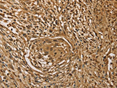
. PRDM14 was detected in paraffin-embedded section of human renal clear cell carcinoma tissue. Heat mediated antigen retrieval was performed in EDTA buffer (pH8.0, epitope retrieval solution). The tissue section was blocked with 10% goat serum. The tissue section was then incubated with 2microg/ml rabbit anti-PRDM14 Antibody (A03870-2) overnight at 4°C. Biotinylated goat anti-rabbit IgG was used as secondary antibody and incubated for 30 minutes at 37°C. The tissue section was developed using Strepavidin-Biotin-Complex (SABC) (Catalog # SA1022) with DAB as the chromogen.")
. PRDM14 was detected in paraffin-embedded section of human rectal cancer tissue. Heat mediated antigen retrieval was performed in EDTA buffer (pH8.0, epitope retrieval solution). The tissue section was blocked with 10% goat serum. The tissue section was then incubated with 2microg/ml rabbit anti-PRDM14 Antibody (A03870-2) overnight at 4°C. Biotinylated goat anti-rabbit IgG was used as secondary antibody and incubated for 30 minutes at 37°C. The tissue section was developed using Strepavidin-Biotin-Complex (SABC) (Catalog # SA1022) with DAB as the chromogen.")
. PRDM14 was detected in immunocytochemical section of HELA cells. Enzyme antigen retrieval was performed using IHC enzyme antigen retrieval reagent (AR0022) for 15 mins. The cells were blocked with 10% goat serum. And then incubated with 5microg/mL rabbit anti-PRDM14 Antibody (A03870-2) overnight at 4°C. DyLight®488 Conjugated Goat Anti-Rabbit IgG (BA1127) was used as secondary antibody at 1:100 dilution and incubated for 30 minutes at 37°C. The section was counterstained with DAPI. Visualize using a fluorescence microscope and filter sets appropriate for the label used.")
Figure 1. Western blot analysis of PRDM14 using anti-PRDM14 antibody (A03870-2). Electrophoresis was performed on a 5-20% SDS-PAGE gel at 70V (Stacking gel) / 90V (Resolving gel) for 2-3 hours. The sample well of each lane was loaded with 30ug of sample under reducing conditions. Lane 1: rat testis tissue lysates, Lane 2: rat spleen tissue lysates, Lane 3: rat C6 whole cell lysates, Lane 4: rat PC-12 whole cell lysates, Lane 5: mouse testis tissue lysates, Lane 6: mouse spleen tissue lysates, Lane 7: mouse NIH/3T3 whole cell lysates, Lane 8: mouse Neuro-2a whole cell lysates. After Electrophoresis, proteins were transferred to a Nitrocellulose membrane at 150mA for 50-90 minutes. Blocked the membrane with 5% Non-fat Milk/ TBS for 1.5 hour at RT. The membrane was incubated with rabbit anti-PRDM14 antigen affinity purified polyclonal antibody (Catalog # A03870-2) at 0.25 microg/mL overnight at 4°C, then washed with TBS-0.1%Tween 3 times with 5 minutes each and probed with a goat anti-rabbit IgG-HRP secondary antibody at a dilution of 1:5000 for 1.5 hour at RT. The signal is developed using an Enhanced Chemiluminescent detection (ECL) kit (Catalog # EK1002) with Tanon 5200 system. A specific band was detected for PRDM14 at approximately 64KD. The expected band size for PRDM14 is at 64KD.
Anti-PRDM14 Antibody Picoband(r)
A03870-2-CARRIER-FREE
ApplicationsFlow Cytometry, ImmunoFluorescence, Western Blot, ImmunoCytoChemistry, ImmunoHistoChemistry
Product group Antibodies
ReactivityHuman, Mouse, Rat
TargetPRDM14
Overview
- SupplierBoster Bio
- Product NameAnti-PRDM14 Antibody Picoband(r)
- Delivery Days Customer9
- ApplicationsFlow Cytometry, ImmunoFluorescence, Western Blot, ImmunoCytoChemistry, ImmunoHistoChemistry
- CertificationResearch Use Only
- ClonalityPolyclonal
- Concentration500 ug/ml
- Gene ID63978
- Target namePRDM14
- Target descriptionPR/SET domain 14
- Target synonymsPFM11, PR domain zinc finger protein 14, PR domain 14, PR domain-containing 14, PR domain-containing protein 14
- HostRabbit
- IsotypeIgG
- Protein IDQ9GZV8
- Protein NamePR domain zinc finger protein 14
- Scientific DescriptionBoster Bio Anti-PRDM14 Antibody Picoband® catalog # A03870-2. Tested in Flow Cytometry, IF, IHC, ICC, WB applications. This antibody reacts with Human, Mouse, Rat. The brand Picoband indicates this is a premium antibody that guarantees superior quality, high affinity, and strong signals with minimal background in Western blot applications. Only our best-performing antibodies are designated as Picoband, ensuring unmatched performance.
- ReactivityHuman, Mouse, Rat
- Storage Instruction-20°C,2°C to 8°C
- UNSPSC12352203
Related products

Product group Antibodies
Anti-PRDM14 AntibodyA84945
ApplicationsWestern Blot, ELISA
ReactivityHuman
- SizePrice
Product group Antibodies

Anti-PRDM14 [RAB-S192]Ab01846-1.1
ApplicationsFlow Cytometry, ImmunoFluorescence
ReactivityHuman
TargetPRDM14
- SizePrice
Product group Antibodies
Anti-PRDM14 Antibody144-60433
ApplicationsImmunoFluorescence, Western Blot, ImmunoHistoChemistry
ReactivityHuman, Mouse, Rat
TargetPRDM14
- SizePrice

Product group Antibodies

Goat anti-PRDM14 (aa414-427)EB10761
ApplicationsFlow Cytometry, ImmunoFluorescence, ELISA
ReactivityBovine, Canine, Human, Mouse, Porcine, Rat
TargetPRDM14
- SizePrice
Product group Antibodies
ApplicationsImmunoPrecipitation, Western Blot, ImmunoCytoChemistry, ImmunoHistoChemistry
ReactivityRat
TargetPRDM14
- SizePrice
Product group Antibodies
PRDM14 AntibodyCSB-PA246357
ApplicationsELISA, ImmunoHistoChemistry
ReactivityHuman, Mouse
TargetPRDM14
- SizePrice
Product group Antibodies

PRDM14 AntibodyLS-C401299
ApplicationsELISA, ImmunoHistoChemistry
ReactivityHuman, Mouse
TargetPRDM14
- SizePrice
![PRDM14 antibody [GT1359] detects PRDM14 protein by Western blot analysis. A. 30 μg 293T whole cell lysate/extract B. 30 μg A431 whole cell lysate/extract C. 30 μg HeLa whole cell lysate/extract D. 30 μg HepG2 whole cell lysate/extract 7.5 % SDS-PAGE PRDM14 antibody [GT1359] (GTX629805) dilution: 1:1000](https://www.genetex.com/upload/website/prouct_img/normal/GTX629805/GTX629805_41456_WB_w_23061202_945.webp)
Product group Antibodies
PRDM14 antibody [GT1359]GTX629805
ApplicationsImmunoFluorescence, Western Blot, ImmunoCytoChemistry
ReactivityHuman
TargetPRDM14
- SizePrice