


Immunohistochemical staining of human testis shows moderate cytoplasmic positivity in acrosomes.
Immunohistochemical staining of human testis shows moderate cytoplasmic positivity in acrosomes.
Anti-PRDM7 Antibody
HPA059944
ApplicationsImmunoHistoChemistry
Product group Antibodies
ReactivityHuman
TargetPRDM7
Overview
- SupplierAtlas Antibodies
- Product NameAnti-PRDM7 Antibody
- Delivery Days Customer4
- ApplicationsImmunoHistoChemistry
- CertificationResearch Use Only
- ClonalityPolyclonal
- ConjugateUnconjugated
- Gene ID11105
- Target namePRDM7
- Target descriptionPR/SET domain 7
- Target synonymsPFM4, ZNF910, histone-lysine N-methyltransferase PRDM7, PR domain 7, PR domain containing 7, PR domain zinc finger protein 7, PR-domain family protein 4, [histone H3]-lysine4 N-methyltransferase PRDM7, probable histone-lysine N-methyltransferase PRDM7
- HostRabbit
- IsotypeIgG
- Protein IDQ9NQW5
- Protein NameHistone-lysine N-methyltransferase PRDM7
- Scientific DescriptionRecombinant Protein Epitope Signature Tag (PrEST) antigen sequence
- ReactivityHuman
- Storage Instruction-20°C,2°C to 8°C
- UNSPSC41116161
Datasheet
MSDS
Related products
Product group Antibodies
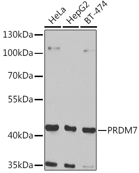
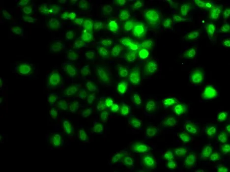
Anti-PRDM7 Antibody144-07961
ApplicationsImmunoFluorescence, Western Blot
ReactivityHuman, Mouse, Rat
TargetPRDM7
- SizePrice

Product group Antibodies
Anti-PRDM7 Antibody Picoband(r)A13923-1-CARRIER-FREE
ApplicationsWestern Blot, ELISA
ReactivityHuman
TargetPRDM7
- SizePrice
Product group Antibodies
PRDM7 AntibodyCSB-PA873626ZA01HU
ApplicationsWestern Blot, ELISA
ReactivityHuman
TargetPRDM7
- SizePrice
Product group Antibodies

PRDM7 AntibodyLS-C409505
ApplicationsImmunoFluorescence, Western Blot
ReactivityHuman, Mouse, Rat
TargetPRDM7
- SizePrice
Product group Antibodies
Anti-PRDM7 AntibodyA16005
ApplicationsImmunoFluorescence, Western Blot, ImmunoCytoChemistry
ReactivityHuman
- SizePrice
Product group Antibodies
Anti-PRDM7Y158473
ApplicationsWestern Blot, ELISA, ImmunoHistoChemistry
ReactivityHuman
- SizePrice
Product group Antibodies
PRDM7 antibodyGTX32808
ApplicationsImmunoFluorescence, Western Blot, ImmunoCytoChemistry
ReactivityHuman
TargetPRDM7
- SizePrice