


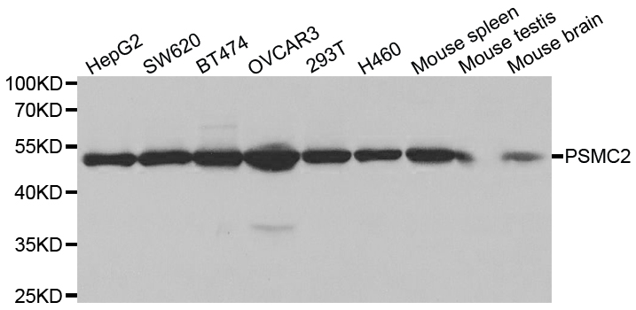
Figure 1. Western blot analysis of PSMC2 using anti-PSMC2 antibody (A09306-1). Electrophoresis was performed on a 5-20% SDS-PAGE gel at 70V (Stacking gel) / 90V (Resolving gel) for 2-3 hours. The sample well of each lane was loaded with 30 ug of sample under reducing conditions. Lane 1: human MOLT-4 whole cell lysates, Lane 2: human T-47D whole cell lysates, Lane 3: monkey COS-7 whole cell lysates. After electrophoresis, proteins were transferred to a nitrocellulose membrane at 150 mA for 50-90 minutes. Blocked the membrane with 5% non-fat milk/TBS for 1.5 hour at RT. The membrane was incubated with rabbit anti-PSMC2 antigen affinity purified polyclonal antibody (Catalog # A09306-1) at 0.5 microg/mL overnight at 4°C, then washed with TBS-0.1%Tween 3 times with 5 minutes each and probed with a goat anti-rabbit IgG-HRP secondary antibody at a dilution of 1:5000 for 1.5 hour at RT. The signal is developed using an Enhanced Chemiluminescent detection (ECL) kit (Catalog # EK1002) with Tanon 5200 system. A specific band was detected for PSMC2 at approximately 49 kDa. The expected band size for PSMC2 is at 49 kDa.
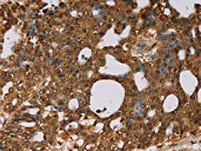
. PSMC2 was detected in an immunocytochemical section of U2OS cells. Enzyme antigen retrieval was performed using IHC enzyme antigen retrieval reagent (AR0022) for 15 mins. The cells were blocked with 10% goat serum. And then incubated with 5 microg/mL rabbit anti-PSMC2 Antibody (A09306-1) overnight at 4°C. Cy3 Conjugated Goat Anti-Rabbit IgG (BA1032) was used as secondary antibody at 1:500 dilution and incubated for 30 minutes at 37°C. The section was counterstained with DAPI. Visualize using a fluorescence microscope and filter sets appropriate for the label used.")
Figure 1. Western blot analysis of PSMC2 using anti-PSMC2 antibody (A09306-1). Electrophoresis was performed on a 5-20% SDS-PAGE gel at 70V (Stacking gel) / 90V (Resolving gel) for 2-3 hours. The sample well of each lane was loaded with 30 ug of sample under reducing conditions. Lane 1: human MOLT-4 whole cell lysates, Lane 2: human T-47D whole cell lysates, Lane 3: monkey COS-7 whole cell lysates. After electrophoresis, proteins were transferred to a nitrocellulose membrane at 150 mA for 50-90 minutes. Blocked the membrane with 5% non-fat milk/TBS for 1.5 hour at RT. The membrane was incubated with rabbit anti-PSMC2 antigen affinity purified polyclonal antibody (Catalog # A09306-1) at 0.5 microg/mL overnight at 4°C, then washed with TBS-0.1%Tween 3 times with 5 minutes each and probed with a goat anti-rabbit IgG-HRP secondary antibody at a dilution of 1:5000 for 1.5 hour at RT. The signal is developed using an Enhanced Chemiluminescent detection (ECL) kit (Catalog # EK1002) with Tanon 5200 system. A specific band was detected for PSMC2 at approximately 49 kDa. The expected band size for PSMC2 is at 49 kDa.
Anti-PSMC2 Antibody Picoband(r)
A09306-1-CARRIER-FREE
ApplicationsImmunoFluorescence, Western Blot, ELISA, ImmunoCytoChemistry
Product group Antibodies
ReactivityHuman, Monkey
TargetPSMC2
Overview
- SupplierBoster Bio
- Product NameAnti-PSMC2 Antibody Picoband(r)
- Delivery Days Customer9
- ApplicationsImmunoFluorescence, Western Blot, ELISA, ImmunoCytoChemistry
- CertificationResearch Use Only
- ClonalityPolyclonal
- Concentration500 ug/ml
- Gene ID5701
- Target namePSMC2
- Target descriptionproteasome 26S subunit, ATPase 2
- Target synonymsMSS1, Nbla10058, RPT1, S7, 26S proteasome regulatory subunit 7, 26S protease regulatory subunit 7, 26S proteasome AAA-ATPase subunit RPT1, mammalian suppressor of sgv-1 of yeast, protease 26S subunit 7, proteasome (prosome, macropain) 26S subunit, ATPase, 2, putative protein product of Nbla10058, testis secretory sperm-binding protein Li 197a
- HostRabbit
- IsotypeIgG
- Protein IDP35998
- Protein Name26S proteasome regulatory subunit 7
- Scientific DescriptionBoster Bio Anti-PSMC2 Antibody Picoband® catalog # A09306-1. Tested in ELISA, IF, ICC, WB applications. This antibody reacts with Human, Monkey. The brand Picoband indicates this is a premium antibody that guarantees superior quality, high affinity, and strong signals with minimal background in Western blot applications. Only our best-performing antibodies are designated as Picoband, ensuring unmatched performance.
- ReactivityHuman, Monkey
- Storage Instruction-20°C,2°C to 8°C
- UNSPSC12352203
Related products
Product group Antibodies
Anti-PSMC2 Antibody144-01985
ApplicationsImmunoFluorescence, Western Blot, ImmunoHistoChemistry
ReactivityHuman, Mouse
TargetPSMC2
- SizePrice
Product group Antibodies
Anti-PSMC2 AntibodyA30543
ApplicationsImmunoFluorescence, Western Blot, ImmunoHistoChemistry
ReactivityHuman, Mouse, Rat
- SizePrice

Product group Antibodies
GTPBP3 Polyclonal AntibodyBS-5120R
ApplicationsImmunoFluorescence, Western Blot, ELISA, ImmunoCytoChemistry, ImmunoHistoChemistry, ImmunoHistoChemistry Frozen, ImmunoHistoChemistry Paraffin
ReactivityChicken, Equine, Human, Mouse, Rat
TargetPSMC2
- SizePrice
Product group Antibodies
PSMC2 AntibodyCSB-PA134529
ApplicationsWestern Blot, ELISA, ImmunoHistoChemistry
ReactivityHuman, Mouse, Rat
TargetPSMC2
- SizePrice
Product group Antibodies
ApplicationsImmunoPrecipitation, Western Blot, ImmunoCytoChemistry, ImmunoHistoChemistry
ReactivityMouse, Rat
TargetPSMC2
- SizePrice
Product group Antibodies

PSMC2 / RPT1 AntibodyLS-C401855
ApplicationsWestern Blot, ELISA, ImmunoHistoChemistry
ReactivityHuman, Mouse, Rat
TargetPSMC2
- SizePrice
Product group Antibodies
PSMC2 antibody, N-termGTX30806
ApplicationsWestern Blot
ReactivityHuman
TargetPSMC2
- SizePrice
Product group Antibodies
Anti-PSMC2 AntibodyHPA019238
ApplicationsWestern Blot, ImmunoHistoChemistry
ReactivityHuman, Mouse, Rat
TargetPSMC2
- SizePrice