Bio-Connect
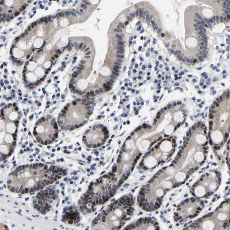
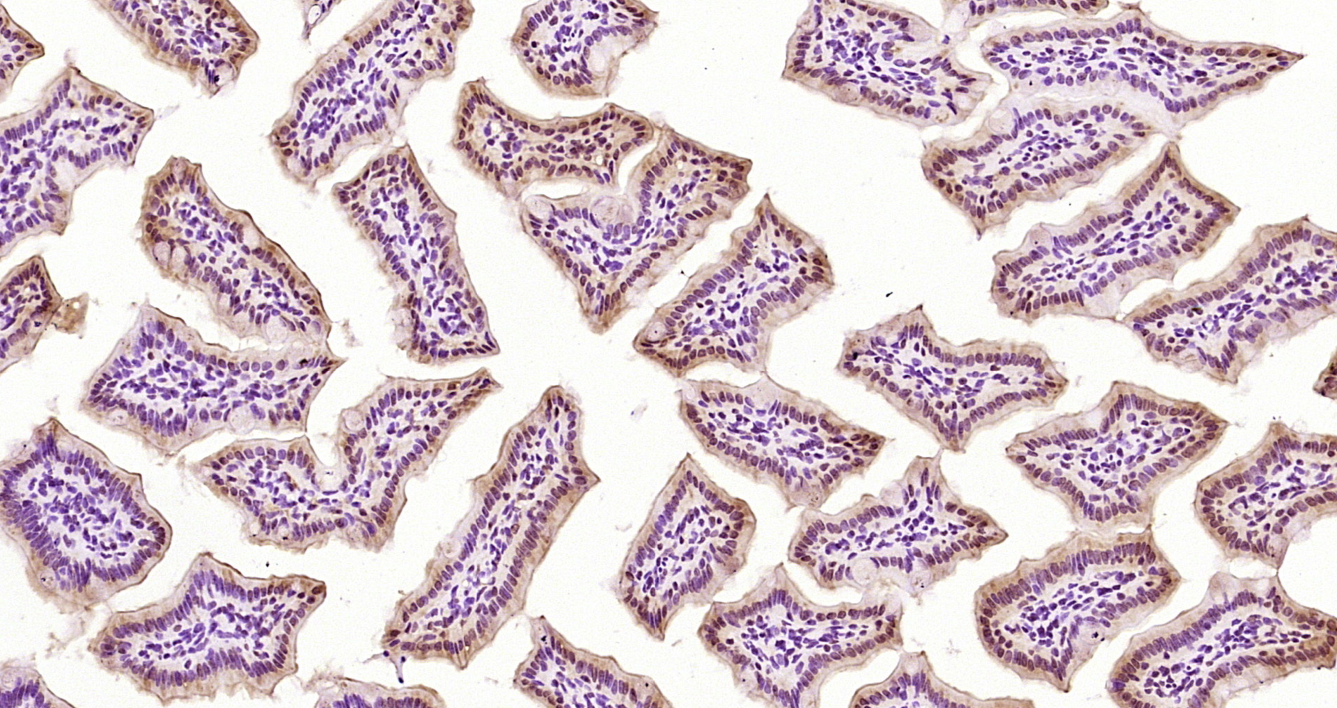
Immunohistochemical staining of human duodenum shows strong nuclear positivity in glandular cells.

Anti-PTRHD1 Antibody

HPA019951
Atlas Antibodies
ApplicationsImmunoCytoChemistry, ImmunoHistoChemistry
Product group Antibodies
ReactivityHuman
TargetPTRHD1
Sign in to order and to see your custom pricing.
Large volume orders?
Order with a bulk request

Overview

  • Supplier
    Atlas Antibodies
  • Product Name
    Anti-PTRHD1
  • Delivery Days Customer
    4
  • Applications
    ImmunoCytoChemistry, ImmunoHistoChemistry
  • Certification
    Research Use Only
  • Clonality
    Polyclonal
  • Conjugate
    Unconjugated
  • Gene ID391356
  • Target name
    PTRHD1
  • Target description
    peptidyl-tRNA hydrolase domain containing 1
  • Target synonyms
    C2orf79; peptidyl-tRNA hydrolase domain-containing protein 1; putative peptidyl-tRNA hydrolase PTRHD1
  • Host
    Rabbit
  • Isotype
    IgG
  • Protein IDQ6GMV3
  • Protein Name
    Putative peptidyl-tRNA hydrolase PTRHD1
  • Scientific Description
    Recombinant Protein Epitope Signature Tag (PrEST) antigen sequence
  • Reactivity
    Human
  • Storage Instruction
    -20°C,2°C to 8°C
  • UNSPSC
    41116161

Related products

Figure 1. Western blot analysis of PTRHD1 using anti-PTRHD1 antibody (A17455). Electrophoresis was performed on a 5-20% SDS-PAGE gel at 70V (Stacking gel) / 90V (Resolving gel) for 2-3 hours. The sample well of each lane was loaded with 30 ug of sample under reducing conditions. Lane 1: human HepG2 whole cell lysates, Lane 2: human 293Tt whole cell lysates, Lane 3: human RT4 whole cell lysates, Lane 4: human A549 whole cell lysates. After electrophoresis, proteins were transferred to a nitrocellulose membrane at 150 mA for 50-90 minutes. Blocked the membrane with 5% non-fat milk/TBS for 1.5 hour at RT. The membrane was incubated with rabbit anti-PTRHD1 antigen affinity purified polyclonal antibody (Catalog # A17455) at 0.5 microg/mL overnight at 4°C, then washed with TBS-0.1%Tween 3 times with 5 minutes each and probed with a goat anti-rabbit IgG-HRP secondary antibody at a dilution of 1:5000 for 1.5 hour at RT. The signal is developed using an Enhanced Chemiluminescent detection (ECL) kit (Catalog # EK1002) with Tanon 5200 system. A specific band was detected for PTRHD1 at approximately 16 kDa. The expected band size for PTRHD1 is at 16 kDa.
Product group Antibodies
Boster Bio
ApplicationsFlow Cytometry, ImmunoFluorescence, Western Blot, ELISA, ImmunoCytoChemistry, ImmunoHistoChemistry
TargetPTRHD1
  • SizePrice
Paraformaldehyde-fixed, paraffin embedded Mouse intestine; Antigen retrieval by boiling in sodium citrate buffer (pH6.0) for 15min; Block endogenous peroxidase by 3% hydrogen peroxide for 20 minutes; Blocking buffer (normal goat serum) at 37°C for 30min; Antibody incubation with PTRHD1 Polyclonal Antibody, Unconjugated (bs-4433R) at 1:200 overnight at 4°C, DAB staining.
Product group Antibodies
Bioss Antibodies
ApplicationsImmunoFluorescence, Western Blot, ELISA, ImmunoCytoChemistry, ImmunoHistoChemistry, ImmunoHistoChemistry Frozen, ImmunoHistoChemistry Paraffin
TargetPTRHD1
  • SizePrice
Atlas Antibodies
Protein IDQ6GMV3
  • SizePrice