


Immunohistochemical staining of human endometrium shows moderate to strong nuclear positivity in glandular cells.

Immunohistochemical staining of human endometrium shows moderate to strong nuclear positivity in glandular cells.
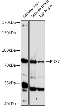
Anti-PUS7 Antibody
HPA024116
ApplicationsWestern Blot
Product group Antibodies
ReactivityHuman, Mouse, Rat
TargetPUS7
Overview
- SupplierAtlas Antibodies
- Product NameAnti-PUS7 Antibody
- Delivery Days Customer4
- ApplicationsWestern Blot
- CertificationResearch Use Only
- ClonalityPolyclonal
- ConjugateUnconjugated
- Gene ID54517
- Target namePUS7
- Target descriptionpseudouridine synthase 7
- Target synonymsIDDABS, pseudouridylate synthase 7 homolog, pseudouridylate synthase 7 (putative)
- HostRabbit
- IsotypeIgG
- Protein IDQ96PZ0
- Protein NamePseudouridylate synthase 7 homolog
- Scientific DescriptionRecombinant Protein Epitope Signature Tag (PrEST) antigen sequence
- ReactivityHuman, Mouse, Rat
- Storage Instruction-20°C,2°C to 8°C
- UNSPSC41116161
Datasheet
MSDS
Related products
Product group Antibodies
PUS7 AntibodyCSB-PA140495XA01SVG
ApplicationsWestern Blot, ELISA
ReactivityYeast
- SizePrice

Product group Antibodies
Anti-PUS7 Antibody Picoband(r)A13434-1-CARRIER-FREE
ApplicationsFlow Cytometry, ImmunoFluorescence, Western Blot, ELISA, ImmunoCytoChemistry, ImmunoHistoChemistry
ReactivityHuman, Mouse, Rat
TargetPUS7
- SizePrice
Product group Antibodies
Anti-PUS7 AntibodyA306343
ApplicationsWestern Blot
ReactivityHuman, Mouse, Rat
- SizePrice
Product group Antibodies

PUS7 Antibody (aa100-150)LS-C762665
ApplicationsWestern Blot
ReactivityHuman
TargetPUS7
- SizePrice
![WB analysis of wild type and PUS7 knock out HeLa cell lysate(10μg per lane) using GTX83755 PUS7 antibody [4A2]. Dilution : 1:500](https://www.genetex.com/upload/website/prouct_img/normal/GTX83755/GTX83755_3885_20181228_WB_w_23061420_122.webp)
Product group Antibodies
PUS7 antibody [4A2]GTX83755
ApplicationsWestern Blot
ReactivityHuman, Monkey
TargetPUS7
- SizePrice