

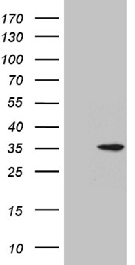
HEK293T cells were transfected with the pCMV6-ENTRY control (Left lane) or pCMV6-ENTRY PRELID1 (Right lane) cDNA for 48 hrs and lysed. Equivalent amounts of cell lysates (5 ug per lane) were separated by SDS-PAGE and immunoblotted with anti-PREL.
 at 120°C for 3min")
HEK293T cells were transfected with the pCMV6-ENTRY control (Left lane) or pCMV6-ENTRY PRELID1 (Right lane) cDNA for 48 hrs and lysed. Equivalent amounts of cell lysates (5 ug per lane) were separated by SDS-PAGE and immunoblotted with anti-PREL.
Anti-PX19 (PRELID1) Mouse Monoclonal Antibody [Clone ID: OTI2A6]
M10248
ApplicationsWestern Blot, ImmunoHistoChemistry
Product group Antibodies
ReactivityHuman, Mouse, Rat
TargetPRELID1
Overview
- SupplierBoster Bio
- Product NameAnti-PX19 (PRELID1) Mouse Monoclonal Antibody [Clone ID: OTI2A6]
- Delivery Days Customer9
- ApplicationsWestern Blot, ImmunoHistoChemistry
- CertificationResearch Use Only
- ClonalityMonoclonal
- Clone IDOTI2A6
- Concentration1 mg/ml
- Gene ID27166
- Target namePRELID1
- Target descriptionPRELI domain containing 1
- Target synonymsCGI-106, PRELI, PX19, SBBI12, PRELI domain-containing protein 1, mitochondrial, 25 kDa protein of relevant evolutionary and lymphoid interest, px19-like protein
- HostMouse
- IsotypeIgG2b
- Protein IDQ9Y255
- Protein NamePRELI domain-containing protein 1, mitochondrial
- Scientific DescriptionBoster Bio PRELID1 mouse monoclonal antibody,clone OTI2A6. Catalog# M10248. Tested in IHC, WB. This antibody reacts with Human, Mouse, Rat.
- ReactivityHuman, Mouse, Rat
- Storage Instruction-20°C
- UNSPSC12352203
Related products
Product group Antibodies
PRELID1 AntibodyCSB-PA018661GA01HU
ApplicationsELISA, ImmunoHistoChemistry
ReactivityHuman, Mouse, Rat
TargetPRELID1
- SizePrice
Product group Antibodies
Anti-PRELID1 AntibodyA45244
ApplicationsImmunoHistoChemistry
ReactivityHuman
- SizePrice
Product group Antibodies

PRELID1 AntibodyLS-C831843
ApplicationsELISA, ImmunoHistoChemistry
ReactivityHuman, Mouse
TargetPRELID1
- SizePrice

Product group Antibodies
Anti-PRELID1 AntibodyHPA005701
ApplicationsImmunoCytoChemistry, ImmunoHistoChemistry
ReactivityHuman
TargetPRELID1
- SizePrice
![PRELID1 antibody [N1C3-2] detects PRELID1 protein at mitochondria by immunofluorescent analysis. Sample: SKNSH cells were fixed in 2% paraformaldehyde/culture medium at 37oC for 30 min. Green: PRELID1 protein stained by PRELID1 antibody [N1C3-2] (GTX118046) diluted at 1:500. Blue: Hoechst 33342 staining. Scale bar = 10 μm.](https://www.genetex.com/upload/website/prouct_img/normal/GTX118046/GTX118046_40590_IFA_w_23060519_552.webp)
Product group Antibodies
PRELID1 antibody [N1C3-2]GTX118046
ApplicationsImmunoFluorescence, Western Blot, ImmunoCytoChemistry, ImmunoHistoChemistry, ImmunoHistoChemistry Paraffin
ReactivityHuman
TargetPRELID1
- SizePrice
Product group Antibodies
Anti-PRELID1Y158462
ApplicationsWestern Blot, ELISA, ImmunoHistoChemistry
ReactivityHuman
- SizePrice