

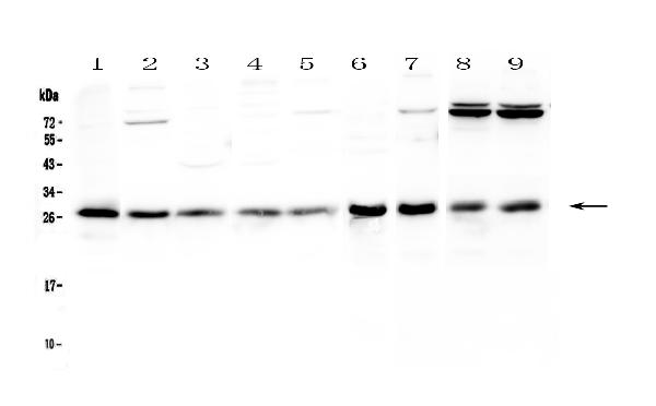
Figure 1. Western blot analysis of RAB27A using anti-RAB27A antibody (A01608-1). Electrophoresis was performed on a 5-20% SDS-PAGE gel at 70V (Stacking gel) / 90V (Resolving gel) for 2-3 hours. The sample well of each lane was loaded with 50ug of sample under reducing conditions. Lane 1: human Hela whole cell lysates, Lane 2: human Jurkat whole cell lysates, Lane 3: human MCF-7 whole cell lysates, Lane 4: human HepG2 whole cell lysates, Lane 5: human A549 whole cell lysates, Lane 6: rat stomach tissue lysates, Lane 7: rat thymus tissue lysates, Lane 8: mouse thymus tissue lysates, Lane 9: mouse NIH3T3 whole cell lysates. After Electrophoresis, proteins were transferred to a Nitrocellulose membrane at 150mA for 50-90 minutes. Blocked the membrane with 5% Non-fat Milk/ TBS for 1.5 hour at RT. The membrane was incubated with rabbit anti-RAB27A antigen affinity purified polyclonal antibody (Catalog # A01608-1) at 0.5 microg/mL overnight at 4°C, then washed with TBS-0.1%Tween 3 times with 5 minutes each and probed with a goat anti-rabbit IgG-HRP secondary antibody at a dilution of 1:10000 for 1.5 hour at RT. The signal is developed using an Enhanced Chemiluminescent detection (ECL) kit (Catalog # EK1002) with Tanon 5200 system. A specific band was detected for RAB27A at approximately 27KD. The expected band size for RAB27A is at 25KD.
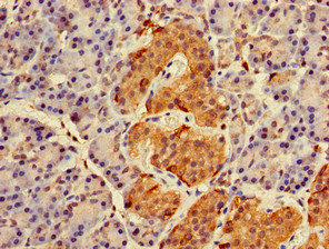
. RAB27A was detected in paraffin-embedded section of human lung cancer tissue. Heat mediated antigen retrieval was performed in citrate buffer (pH6, epitope retrieval solution) for 20 mins. The tissue section was blocked with 10% goat serum. The tissue section was then incubated with 1microg/ml rabbit anti-RAB27A Antibody (A01608-1) overnight at 4°C. Biotinylated goat anti-rabbit IgG was used as secondary antibody and incubated for 30 minutes at 37°C. The tissue section was developed using Strepavidin-Biotin-Complex (SABC)(Catalog # SA1022) with DAB as the chromogen.")
. RAB27A was detected in paraffin-embedded section of human mammary cancer tissue. Heat mediated antigen retrieval was performed in citrate buffer (pH6, epitope retrieval solution) for 20 mins. The tissue section was blocked with 10% goat serum. The tissue section was then incubated with 1microg/ml rabbit anti-RAB27A Antibody (A01608-1) overnight at 4°C. Biotinylated goat anti-rabbit IgG was used as secondary antibody and incubated for 30 minutes at 37°C. The tissue section was developed using Strepavidin-Biotin-Complex (SABC)(Catalog # SA1022) with DAB as the chromogen.")
. RAB27A was detected in paraffin-embedded section of rat small intestine tissue. Heat mediated antigen retrieval was performed in citrate buffer (pH6, epitope retrieval solution) for 20 mins. The tissue section was blocked with 10% goat serum. The tissue section was then incubated with 1microg/ml rabbit anti-RAB27A Antibody (A01608-1) overnight at 4°C. Biotinylated goat anti-rabbit IgG was used as secondary antibody and incubated for 30 minutes at 37°C. The tissue section was developed using Strepavidin-Biotin-Complex (SABC)(Catalog # SA1022) with DAB as the chromogen.")
. RAB27A was detected in paraffin-embedded section of rat spleen tissue. Heat mediated antigen retrieval was performed in citrate buffer (pH6, epitope retrieval solution) for 20 mins. The tissue section was blocked with 10% goat serum. The tissue section was then incubated with 1microg/ml rabbit anti-RAB27A Antibody (A01608-1) overnight at 4°C. Biotinylated goat anti-rabbit IgG was used as secondary antibody and incubated for 30 minutes at 37°C. The tissue section was developed using Strepavidin-Biotin-Complex (SABC)(Catalog # SA1022) with DAB as the chromogen.")
. RAB27A was detected in paraffin-embedded section of mouse spleen tissue. Heat mediated antigen retrieval was performed in citrate buffer (pH6, epitope retrieval solution) for 20 mins. The tissue section was blocked with 10% goat serum. The tissue section was then incubated with 1microg/ml rabbit anti-RAB27A Antibody (A01608-1) overnight at 4°C. Biotinylated goat anti-rabbit IgG was used as secondary antibody and incubated for 30 minutes at 37°C. The tissue section was developed using Strepavidin-Biotin-Complex (SABC)(Catalog # SA1022) with DAB as the chromogen.")
. Overlay histogram showing K562 cells stained with A01608-1 (Blue line). To facilitate intracellular staining, cells were fixed with 4% paraformaldehyde and permeabilized with permeabilization buffer. The cells were blocked with 10% normal goat serum. And then incubated with rabbit anti-RAB27A Antibody (A01608-1,1microg/1x106 cells) for 30 min at 20°C. DyLight®488 conjugated goat anti-rabbit IgG (BA1127, 5-10microg/1x106 cells) was used as secondary antibody for 30 minutes at 20°C. Isotype control antibody (Green line) was rabbit IgG (1microg/1x106) used under the same conditions. Unlabelled sample without incubation with primary antibody and secondary antibody (Red line) was used as a blank control.")
Figure 1. Western blot analysis of RAB27A using anti-RAB27A antibody (A01608-1). Electrophoresis was performed on a 5-20% SDS-PAGE gel at 70V (Stacking gel) / 90V (Resolving gel) for 2-3 hours. The sample well of each lane was loaded with 50ug of sample under reducing conditions. Lane 1: human Hela whole cell lysates, Lane 2: human Jurkat whole cell lysates, Lane 3: human MCF-7 whole cell lysates, Lane 4: human HepG2 whole cell lysates, Lane 5: human A549 whole cell lysates, Lane 6: rat stomach tissue lysates, Lane 7: rat thymus tissue lysates, Lane 8: mouse thymus tissue lysates, Lane 9: mouse NIH3T3 whole cell lysates. After Electrophoresis, proteins were transferred to a Nitrocellulose membrane at 150mA for 50-90 minutes. Blocked the membrane with 5% Non-fat Milk/ TBS for 1.5 hour at RT. The membrane was incubated with rabbit anti-RAB27A antigen affinity purified polyclonal antibody (Catalog # A01608-1) at 0.5 microg/mL overnight at 4°C, then washed with TBS-0.1%Tween 3 times with 5 minutes each and probed with a goat anti-rabbit IgG-HRP secondary antibody at a dilution of 1:10000 for 1.5 hour at RT. The signal is developed using an Enhanced Chemiluminescent detection (ECL) kit (Catalog # EK1002) with Tanon 5200 system. A specific band was detected for RAB27A at approximately 27KD. The expected band size for RAB27A is at 25KD.
Anti-RAB27A Antibody Picoband(r)
A01608-1-CARRIER-FREE
ApplicationsFlow Cytometry, Western Blot, ELISA, ImmunoCytoChemistry, ImmunoHistoChemistry
Product group Antibodies
ReactivityHuman, Mouse, Rat
TargetRAB27A
Overview
- SupplierBoster Bio
- Product NameAnti-RAB27A Antibody Picoband(r)
- Delivery Days Customer9
- ApplicationsFlow Cytometry, Western Blot, ELISA, ImmunoCytoChemistry, ImmunoHistoChemistry
- CertificationResearch Use Only
- ClonalityPolyclonal
- Concentration500 ug/ml
- Gene ID5873
- Target nameRAB27A
- Target descriptionRAB27A, member RAS oncogene family
- Target synonymsGS2, HsT18676, RAB27, RAM, ras-related protein Rab-27A, GTP-binding protein Ram, mutant Ras-related protein Rab-27A, rab-27
- HostRabbit
- IsotypeIgG
- Protein IDP51159
- Protein NameRas-related protein Rab-27A
- Scientific DescriptionBoster Bio Anti-RAB27A Antibody Picoband® catalog # A01608-1. Tested in ELISA, Flow Cytometry, IHC, ICC, WB applications. This antibody reacts with Human, Mouse, Rat. The brand Picoband indicates this is a premium antibody that guarantees superior quality, high affinity, and strong signals with minimal background in Western blot applications. Only our best-performing antibodies are designated as Picoband, ensuring unmatched performance.
- ReactivityHuman, Mouse, Rat
- Storage Instruction-20°C,2°C to 8°C
- UNSPSC12352203
Related products
Product group Antibodies
Anti-RAB27A AntibodyA121563
ApplicationsImmunoFluorescence, Western Blot, ImmunoHistoChemistry, ImmunoHistoChemistry Frozen, ImmunoHistoChemistry Paraffin
ReactivityCanine, Human, Monkey, Mouse, Rat
- SizePrice
Product group Antibodies
Anti-RAB27A Antibody144-01934
ApplicationsImmunoFluorescence, Western Blot, ImmunoHistoChemistry
ReactivityHuman, Mouse
TargetRAB27A
- SizePrice
Product group Antibodies
Rab27a Recombinant Antibody, AbBy Fluor-647 ConjugatedBSM-62025R-BF647
ApplicationsWestern Blot
ReactivityHuman
TargetRAB27A
- SizePrice
Product group Antibodies
Rab27A Polyclonal AntibodyCAC08772
ApplicationsImmunoFluorescence, ELISA, ImmunoHistoChemistry
TargetRAB27A
- SizePrice
Product group Antibodies
RAB27A AntibodyCSB-PA019177LA01HU
ApplicationsImmunoFluorescence, ELISA, ImmunoHistoChemistry
ReactivityHuman
TargetRAB27A
- SizePrice
Product group Antibodies

RAB27A / RAB27 AntibodyLS-C401961
ApplicationsWestern Blot, ELISA, ImmunoHistoChemistry
ReactivityHuman, Mouse, Rat
TargetRAB27A
- SizePrice

Product group Antibodies
Anti-RAB27A AntibodyHPA001333
ApplicationsWestern Blot, ImmunoHistoChemistry
ReactivityHuman
TargetRAB27A
- SizePrice
Product group Antibodies
RAB27A antibody [N1C3]GTX109180
ApplicationsWestern Blot, ImmunoHistoChemistry, ImmunoHistoChemistry Paraffin
ReactivityHuman
TargetRAB27A
- SizePrice
Product group Antibodies
Anti-RAB27AY158078
ApplicationsWestern Blot, ELISA, ImmunoHistoChemistry
ReactivityHuman, Mouse, Rat
- SizePrice