


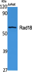
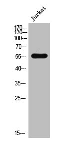
Figure 1. Western blot analysis of RAD18 using anti-RAD18 antibody (A01622-2). Electrophoresis was performed on a 5-20% SDS-PAGE gel at 70V (Stacking gel) / 90V (Resolving gel) for 2-3 hours. The sample well of each lane was loaded with 30 ug of sample under reducing conditions. Lane 1: human Hela whole cell lysates, Lane 2: human 293T whole cell lysates, Lane 3: human K562 whole cell lysates, Lane 4: rat C6 whole cell lysates, Lane 5: mouse NIH/3T3 whole cell lysates. After electrophoresis, proteins were transferred to a nitrocellulose membrane at 150 mA for 50-90 minutes. Blocked the membrane with 5% non-fat milk/TBS for 1.5 hour at RT. The membrane was incubated with rabbit anti-RAD18 antigen affinity purified polyclonal antibody (Catalog # A01622-2) at 0.5 microg/mL overnight at 4°C, then washed with TBS-0.1%Tween 3 times with 5 minutes each and probed with a goat anti-rabbit IgG-HRP secondary antibody at a dilution of 1:5000 for 1.5 hour at RT. The signal is developed using an Enhanced Chemiluminescent detection (ECL) kit (Catalog # EK1002) with Tanon 5200 system. A specific band was detected for RAD18 at approximately 75 kDa. The expected band size for RAD18 is at 56 kDa.
. RAD18 was detected in a paraffin-embedded section of human gastric signet ring cell carcinoma tissue. Heat mediated antigen retrieval was performed in EDTA buffer (pH 8.0, epitope retrieval solution). The tissue section was blocked with 10% goat serum. The tissue section was then incubated with 2 microg/ml rabbit anti-RAD18 Antibody (A01622-2) overnight at 4°C. Biotinylated goat anti-rabbit IgG was used as secondary antibody and incubated for 30 minutes at 37°C. The tissue section was developed using Strepavidin-Biotin-Complex (SABC) (Catalog # SA1022) with DAB as the chromogen.")
. RAD18 was detected in a paraffin-embedded section of squamous metaplasia of human renal pelvis tissue. Heat mediated antigen retrieval was performed in EDTA buffer (pH 8.0, epitope retrieval solution). The tissue section was blocked with 10% goat serum. The tissue section was then incubated with 2 microg/ml rabbit anti-RAD18 Antibody (A01622-2) overnight at 4°C. Biotinylated goat anti-rabbit IgG was used as secondary antibody and incubated for 30 minutes at 37°C. The tissue section was developed using Strepavidin-Biotin-Complex (SABC) (Catalog # SA1022) with DAB as the chromogen.")
. RAD18 was detected in a paraffin-embedded section of human tonsil tissue. Heat mediated antigen retrieval was performed in EDTA buffer (pH 8.0, epitope retrieval solution). The tissue section was blocked with 10% goat serum. The tissue section was then incubated with 2 microg/ml rabbit anti-RAD18 Antibody (A01622-2) overnight at 4°C. Biotinylated goat anti-rabbit IgG was used as secondary antibody and incubated for 30 minutes at 37°C. The tissue section was developed using Strepavidin-Biotin-Complex (SABC) (Catalog # SA1022) with DAB as the chromogen.")
. Overlay histogram showing K562 cells stained with A01622-2 (Blue line). To facilitate intracellular staining, cells were fixed with 4% paraformaldehyde and permeabilized with permeabilization buffer. The cells were blocked with 10% normal goat serum. And then incubated with rabbit anti-RAD18 Antibody (A01622-2, 1 microg/1x106 cells) for 30 min at 20°C. DyLight®488 conjugated goat anti-rabbit IgG (BA1127, 5-10 microg/1x106 cells) was used as secondary antibody for 30 minutes at 20°C. Isotype control antibody (Green line) was rabbit IgG (1 microg/1x106) used under the same conditions. Unlabelled sample without incubation with primary antibody and secondary antibody (Red line) was used as a blank control.")
Figure 1. Western blot analysis of RAD18 using anti-RAD18 antibody (A01622-2). Electrophoresis was performed on a 5-20% SDS-PAGE gel at 70V (Stacking gel) / 90V (Resolving gel) for 2-3 hours. The sample well of each lane was loaded with 30 ug of sample under reducing conditions. Lane 1: human Hela whole cell lysates, Lane 2: human 293T whole cell lysates, Lane 3: human K562 whole cell lysates, Lane 4: rat C6 whole cell lysates, Lane 5: mouse NIH/3T3 whole cell lysates. After electrophoresis, proteins were transferred to a nitrocellulose membrane at 150 mA for 50-90 minutes. Blocked the membrane with 5% non-fat milk/TBS for 1.5 hour at RT. The membrane was incubated with rabbit anti-RAD18 antigen affinity purified polyclonal antibody (Catalog # A01622-2) at 0.5 microg/mL overnight at 4°C, then washed with TBS-0.1%Tween 3 times with 5 minutes each and probed with a goat anti-rabbit IgG-HRP secondary antibody at a dilution of 1:5000 for 1.5 hour at RT. The signal is developed using an Enhanced Chemiluminescent detection (ECL) kit (Catalog # EK1002) with Tanon 5200 system. A specific band was detected for RAD18 at approximately 75 kDa. The expected band size for RAD18 is at 56 kDa.
Anti-RAD18 Antibody Picoband(r)
A01622-2-CY3
ApplicationsFlow Cytometry, Western Blot, ELISA, ImmunoHistoChemistry
Product group Antibodies
ReactivityHuman, Mouse, Rat
TargetRAD18
Overview
- SupplierBoster Bio
- Product NameAnti-RAD18 Antibody Picoband(r)
- Delivery Days Customer9
- ApplicationsFlow Cytometry, Western Blot, ELISA, ImmunoHistoChemistry
- CertificationResearch Use Only
- ClonalityPolyclonal
- Concentration500 ug/ml
- ConjugateCy3
- Gene ID56852
- Target nameRAD18
- Target descriptionRAD18 E3 ubiquitin protein ligase
- Target synonymsRNF73, E3 ubiquitin-protein ligase RAD18, RAD18 homolog, RAD18, S. cerevisiae, homolog, RING finger protein 73, RING-type E3 ubiquitin transferase RAD18, hHR18, hRAD18, postreplication repair protein RAD18, postreplication repair protein hRAD18p
- HostRabbit
- IsotypeIgG
- Protein IDQ9NS91
- Protein NameE3 ubiquitin-protein ligase RAD18
- Scientific DescriptionBoster Bio Anti-RAD18 Antibody Picoband® catalog # A01622-2. Tested in ELISA, Flow Cytometry, IHC, WB applications. This antibody reacts with Human, Mouse, Rat. The brand Picoband indicates this is a premium antibody that guarantees superior quality, high affinity, and strong signals with minimal background in Western blot applications. Only our best-performing antibodies are designated as Picoband, ensuring unmatched performance.
- ReactivityHuman, Mouse, Rat
- Storage Instruction-20°C,2°C to 8°C
- UNSPSC12352203
Related products
Product group Antibodies
RAD18 Polyclonal AntibodyCAC14491
ApplicationsWestern Blot, ELISA, ImmunoHistoChemistry
TargetRAD18
- SizePrice
Product group Antibodies
Anti-RAD18 Antibody144-05380
ApplicationsWestern Blot
ReactivityHuman, Mouse
TargetRAD18
- SizePrice
Product group Antibodies
Anti-RAD18 AntibodyA99603
ApplicationsWestern Blot, ELISA
ReactivityHuman, Rat
- SizePrice
Product group Antibodies
Anti-RAD18 Antibody Picoband(r)A01622-2-CARRIER-FREE
ApplicationsFlow Cytometry, Western Blot, ELISA, ImmunoHistoChemistry
ReactivityHuman, Mouse, Rat
TargetRAD18
- SizePrice
Product group Antibodies
RAD18 AntibodyCSB-PA003913
ApplicationsWestern Blot, ELISA
ReactivityHuman, Rat
TargetRAD18
- SizePrice
Product group Antibodies
RAD18 antibody [N1C1]GTX113987
ApplicationsImmunoFluorescence, ImmunoCytoChemistry, ImmunoHistoChemistry, ImmunoHistoChemistry Paraffin
ReactivityHuman
TargetRAD18
- SizePrice
Product group Antibodies

RAD18 AntibodyLS-C748484
ApplicationsImmunoFluorescence, Western Blot, ImmunoHistoChemistry
ReactivityHuman, Mouse, Rat
TargetRAD18
- SizePrice
Product group Antibodies
RAD18 Recombinant AntibodyBSM-61274R
ApplicationsFlow Cytometry, Western Blot
TargetRAD18
- SizePrice