


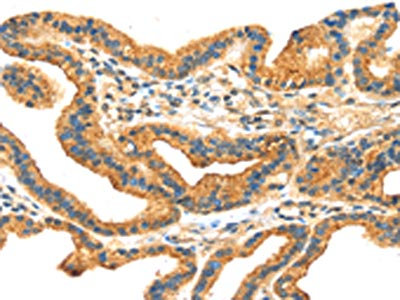
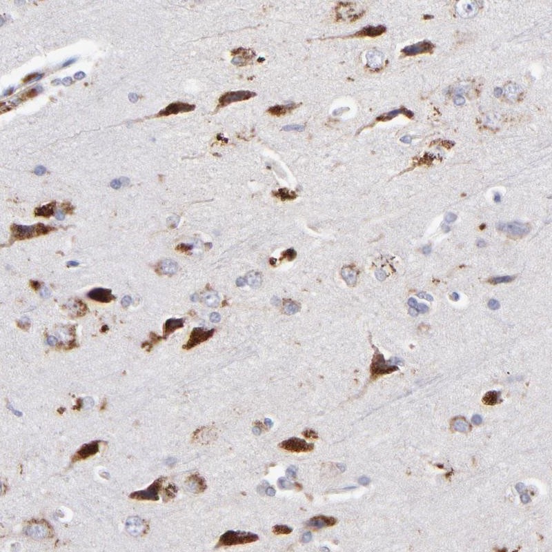
Figure 3. Immunohistochemistry validation of RRAGA using Anti-Rag A/B RRAGA Antibody (A06883-1). Immunohistochemical analysis of paraffin-embedded Human brain using Anti-Rag A/B polyclonal antibody. Antibody was diluted at 1:100 (4°C
. Immunohistochemical analysis of paraffin-embedded Human brain using Anti-Rag A/B polyclonal antibody. Antibody was diluted at 1:100 (4°C")
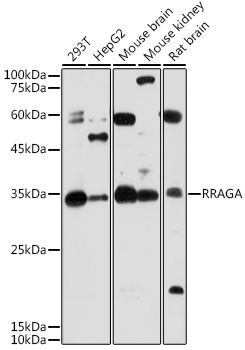
 analysis of mouse brain cells using Anti-Rag A/B polyclonal antibody. Electrophoresis was performed on a SDS-PAGE gel. To determine SDS-PAGE gel concentration")
 analysis of HepG2 cells using Anti-Rag A/B polyclonal antibody. Electrophoresis was performed on a SDS-PAGE gel. To determine SDS-PAGE gel concentration")
Figure 3. Immunohistochemistry validation of RRAGA using Anti-Rag A/B RRAGA Antibody (A06883-1). Immunohistochemical analysis of paraffin-embedded Human brain using Anti-Rag A/B polyclonal antibody. Antibody was diluted at 1:100 (4°C
Anti-Rag A/B RRAGA Antibody
A06883-1
ApplicationsImmunoFluorescence, Western Blot, ELISA, ImmunoHistoChemistry
Product group Antibodies
ReactivityHuman, Mouse, Rat
TargetRRAGA
Overview
- SupplierBoster Bio
- Product NameAnti-Rag A/B RRAGA Antibody
- Delivery Days Customer9
- ApplicationsImmunoFluorescence, Western Blot, ELISA, ImmunoHistoChemistry
- CertificationResearch Use Only
- ClonalityPolyclonal
- Concentration1 mg/ml
- Gene ID10670
- Target nameRRAGA
- Target descriptionRas related GTP binding A
- Target synonymsFIP-1, FIP1, RAGA, ras-related GTP-binding protein A, adenovirus E3 14.7 kDa-interacting protein 1, adenovirus E3-14.7K interacting protein 1, rag A
- HostRabbit
- IsotypeIgG
- Protein IDQ7L523
- Protein NameRas-related GTP-binding protein A
- Scientific DescriptionBoster Bio Anti-Rag A/B RRAGA Antibody catalog # A06883-1. Tested in WB, IHC, IF, ELISA applications. This antibody reacts with Human, Mouse, Rat.
- ReactivityHuman, Mouse, Rat
- Storage Instruction-20°C,2°C to 8°C
- UNSPSC12352203
Related products
Product group Antibodies
RRAGA AntibodyCSB-PA022239
ApplicationsELISA, ImmunoHistoChemistry
ReactivityHuman, Mouse, Rat
TargetRRAGA
- SizePrice
Product group Antibodies
Anti-Rag A AntibodyA15885
ApplicationsImmunoFluorescence, Western Blot, ImmunoCytoChemistry
ReactivityHuman, Mouse, Rat
- SizePrice
Product group Antibodies
Anti-RRAGA Antibody144-63597
ApplicationsWestern Blot
ReactivityHuman, Mouse, Rat
TargetRRAGA
- SizePrice
Product group Antibodies
Anti-RRAGA AntibodyHPA003734
ApplicationsWestern Blot, ImmunoCytoChemistry, ImmunoHistoChemistry
ReactivityHuman
TargetRRAGA
- SizePrice
Product group Antibodies

RRAGA AntibodyLS-C405314
ApplicationsWestern Blot, ELISA, ImmunoHistoChemistry
ReactivityHuman, Mouse, Rat
TargetRRAGA
- SizePrice
Product group Antibodies
RRAGA + RRAGB Polyclonal AntibodyBS-10731R
ApplicationsImmunoFluorescence, ELISA, ImmunoCytoChemistry, ImmunoHistoChemistry, ImmunoHistoChemistry Frozen, ImmunoHistoChemistry Paraffin
ReactivityCanine, Equine, Human, Mouse, Porcine, Rat, Sheep
TargetRRAGA
- SizePrice
![Rag A antibody detects Rag A protein at cytoplasm and nucleus by immunofluorescent analysis. Sample: HeLa cells were fixed in 4% paraformaldehyde at RT for 15 min. Green: Rag A stained by Rag A antibody (GTX135318) diluted at 1:500. Red: alpha Tubulin, a cytoskeleton marker, stained by alpha Tubulin antibody [GT114] (GTX628802) diluted at 1:1000. Scale bar= 10μm.](https://www.genetex.com/upload/website/prouct_img/normal/GTX135318/GTX135318_44447_20220408_ICC_IF_w_23051500_990.webp)
Product group Antibodies
Rag A antibodyGTX135318
ApplicationsImmunoFluorescence, Western Blot, ImmunoCytoChemistry, ImmunoHistoChemistry, ImmunoHistoChemistry Paraffin
ReactivityHuman, Mouse, Rat
TargetRRAGA
- SizePrice