


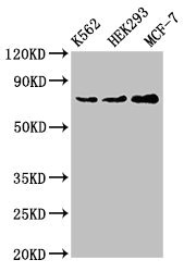
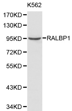
Figure 1. Western blot analysis of RALBP1 using anti-RALBP1 antibody (M01403-4). Electrophoresis was performed on a 5-20% SDS-PAGE gel at 70V (Stacking gel) / 90V (Resolving gel) for 2-3 hours. The sample well of each lane was loaded with 30 ug of sample under reducing conditions. Lane 1: human HL-60 whole cell lysates, Lane 2: human Daudi whole cell lysates, Lane 3: human SW620 whole cell lysates, Lane 4: rat brain tissue lysates, Lane 5: mouse lung tissue lysates, Lane 6: mouse brain tissue lysates. After electrophoresis, proteins were transferred to a nitrocellulose membrane at 150 mA for 50-90 minutes. Blocked the membrane with 5% non-fat milk/TBS for 1.5 hour at RT. The membrane was incubated with mouse anti-RALBP1 antigen affinity purified monoclonal antibody (Catalog # M01403-4) at 0.5 microg/mL overnight at 4°C, then washed with TBS-0.1%Tween 3 times with 5 minutes each and probed with a goat anti-mouse IgG-HRP secondary antibody at a dilution of 1:10000 for 1.5 hour at RT. The signal is developed using an Enhanced Chemiluminescent detection (ECL) kit (Catalog # EK1001) with Tanon 5200 system. A specific band was detected for RALBP1 at approximately 95 kDa. The expected band size for RALBP1 is at 95 kDa.
. RALBP1 was detected in a paraffin-embedded section of human ovarian cancer tissue. Heat mediated antigen retrieval was performed in EDTA buffer (pH 8.0, epitope retrieval solution). The tissue section was blocked with 10% goat serum. The tissue section was then incubated with 2 microg/ml mouse anti-RALBP1 Antibody (M01403-4) overnight at 4°C. Biotinylated goat anti-mouse IgG was used as secondary antibody and incubated for 30 minutes at 37°C. The tissue section was developed using Strepavidin-Biotin-Complex (SABC) (Catalog # SA1021) with DAB as the chromogen.")
. RALBP1 was detected in a paraffin-embedded section of human rectum adenocarcinoma tissue. Heat mediated antigen retrieval was performed in EDTA buffer (pH 8.0, epitope retrieval solution). The tissue section was blocked with 10% goat serum. The tissue section was then incubated with 2 microg/ml mouse anti-RALBP1 Antibody (M01403-4) overnight at 4°C. Biotinylated goat anti-mouse IgG was used as secondary antibody and incubated for 30 minutes at 37°C. The tissue section was developed using Strepavidin-Biotin-Complex (SABC) (Catalog # SA1021) with DAB as the chromogen.")
. RALBP1 was detected in a paraffin-embedded section of human lung cancer tissue. Heat mediated antigen retrieval was performed in EDTA buffer (pH 8.0, epitope retrieval solution). The tissue section was blocked with 10% goat serum. The tissue section was then incubated with 2 microg/ml mouse anti-RALBP1 Antibody (M01403-4) overnight at 4°C. Biotinylated goat anti-mouse IgG was used as secondary antibody and incubated for 30 minutes at 37°C. The tissue section was developed using Strepavidin-Biotin-Complex (SABC) (Catalog # SA1021) with DAB as the chromogen.")
. RALBP1 was detected in a paraffin-embedded section of human metaplasia of squamous cells of the renal pelvis tissue. Heat mediated antigen retrieval was performed in EDTA buffer (pH 8.0, epitope retrieval solution). The tissue section was blocked with 10% goat serum. The tissue section was then incubated with 2 microg/ml mouse anti-RALBP1 Antibody (M01403-4) overnight at 4°C. Biotinylated goat anti-mouse IgG was used as secondary antibody and incubated for 30 minutes at 37°C. The tissue section was developed using Strepavidin-Biotin-Complex (SABC) (Catalog # SA1021) with DAB as the chromogen.")
. RALBP1 was detected in a paraffin-embedded section of mouse brain tissue. Heat mediated antigen retrieval was performed in EDTA buffer (pH 8.0, epitope retrieval solution). The tissue section was blocked with 10% goat serum. The tissue section was then incubated with 2 microg/ml mouse anti-RALBP1 Antibody (M01403-4) overnight at 4°C. Biotinylated goat anti-mouse IgG was used as secondary antibody and incubated for 30 minutes at 37°C. The tissue section was developed using Strepavidin-Biotin-Complex (SABC) (Catalog # SA1021) with DAB as the chromogen.")
. RALBP1 was detected in an immunocytochemical section of U20S cells. Enzyme antigen retrieval was performed using IHC enzyme antigen retrieval reagent (AR0022) for 15 mins. The cells were blocked with 10% goat serum. And then incubated with 5 microg/mL mouse anti-RALBP1 Antibody (M01403-4) overnight at 4°C. DyLight®488 Conjugated Goat Anti-Mouse IgG (BA1126) was used as secondary antibody at 1:100 dilution and incubated for 30 minutes at 37°C. The section was counterstained with DAPI. Visualize using a fluorescence microscope and filter sets appropriate for the label used.")
. Overlay histogram showing SiHa cells stained with M01403-4 (Blue line). To facilitate intracellular staining, cells were fixed with 4% paraformaldehyde and permeabilized with permeabilization buffer. The cells were blocked with 10% normal goat serum. And then incubated with mouse anti-RALBP1 Antibody (M01403-4, 1 microg/1x106 cells) for 30 min at 20°C. DyLight®488 conjugated goat anti-mouse IgG (BA1126, 5-10 microg/1x106 cells) was used as secondary antibody for 30 minutes at 20°C. Isotype control antibody (Green line) was mouse IgG (1 microg/1x106) used under the same conditions. Unlabelled sample without incubation with primary antibody and secondary antibody (Red line) was used as a blank control.")
Figure 1. Western blot analysis of RALBP1 using anti-RALBP1 antibody (M01403-4). Electrophoresis was performed on a 5-20% SDS-PAGE gel at 70V (Stacking gel) / 90V (Resolving gel) for 2-3 hours. The sample well of each lane was loaded with 30 ug of sample under reducing conditions. Lane 1: human HL-60 whole cell lysates, Lane 2: human Daudi whole cell lysates, Lane 3: human SW620 whole cell lysates, Lane 4: rat brain tissue lysates, Lane 5: mouse lung tissue lysates, Lane 6: mouse brain tissue lysates. After electrophoresis, proteins were transferred to a nitrocellulose membrane at 150 mA for 50-90 minutes. Blocked the membrane with 5% non-fat milk/TBS for 1.5 hour at RT. The membrane was incubated with mouse anti-RALBP1 antigen affinity purified monoclonal antibody (Catalog # M01403-4) at 0.5 microg/mL overnight at 4°C, then washed with TBS-0.1%Tween 3 times with 5 minutes each and probed with a goat anti-mouse IgG-HRP secondary antibody at a dilution of 1:10000 for 1.5 hour at RT. The signal is developed using an Enhanced Chemiluminescent detection (ECL) kit (Catalog # EK1001) with Tanon 5200 system. A specific band was detected for RALBP1 at approximately 95 kDa. The expected band size for RALBP1 is at 95 kDa.
Anti-RALBP1 Antibody Picoband(r) (monoclonal, 2F9D3)
M01403-4-DYLIGHT594
ApplicationsFlow Cytometry, ImmunoFluorescence, Western Blot, ImmunoCytoChemistry, ImmunoHistoChemistry
Product group Antibodies
TargetRALBP1
Overview
- SupplierBoster Bio
- Product NameAnti-RALBP1 Antibody Picoband(r) (monoclonal, 2F9D3)
- Delivery Days Customer9
- ApplicationsFlow Cytometry, ImmunoFluorescence, Western Blot, ImmunoCytoChemistry, ImmunoHistoChemistry
- CertificationResearch Use Only
- ClonalityMonoclonal
- Clone ID2F9D3
- Concentration500 ug/ml
- ConjugateOther Conjugate
- Gene ID10928
- Target nameRALBP1
- Target descriptionralA binding protein 1
- Target synonyms76 kDa Ral-interacting protein; dinitrophenyl S-glutathione ATPase; DNP-SG ATPase; ralA-binding protein 1; ral-interacting protein 1; RIP1; RLIP1; RLIP76
- HostMouse
- IsotypeIgG2b
- Protein IDQ15311
- Protein NameRalA-binding protein 1
- Scientific DescriptionBoster Bio Anti-RALBP1 Antibody Picoband® (monoclonal, 2F9D3) catalog # M01403-4. Tested in Flow Cytometry, IF, IHC, ICC, WB applications. This antibody reacts with Human, Mouse, Rat. The brand Picoband indicates this is a premium antibody that guarantees superior quality, high affinity, and strong signals with minimal background in Western blot applications. Only our best-performing antibodies are designated as Picoband, ensuring unmatched performance.
- Storage Instruction-20°C,2°C to 8°C
- UNSPSC12352203
Related products
Product group Antibodies
Anti-RALBP1 Antibody144-01140
ApplicationsWestern Blot, ImmunoHistoChemistry
TargetRALBP1
- SizePrice
Product group Antibodies
ApplicationsImmunoPrecipitation
TargetRALBP1
- SizePrice
Product group Antibodies
RALBP1 Polyclonal AntibodyCAC14895
ApplicationsImmunoFluorescence, Western Blot, ELISA, ImmunoHistoChemistry
TargetRALBP1
- SizePrice
Product group Antibodies
RALBP1 Recombinant AntibodyBSM-61265R
ApplicationsFlow Cytometry, ImmunoFluorescence, Western Blot, ImmunoHistoChemistry, ImmunoHistoChemistry Frozen, ImmunoHistoChemistry Paraffin
TargetRALBP1
- SizePrice
Product group Antibodies
RALBP1 AntibodyCSB-PA618981LA01HU
ApplicationsImmunoFluorescence, Western Blot, ELISA, ImmunoHistoChemistry
ReactivityHuman
TargetRALBP1
- SizePrice

Product group Antibodies
Anti-RALBP1 AntibodyHPA007622
ApplicationsImmunoCytoChemistry
ReactivityHuman
TargetRALBP1
- SizePrice

Product group Antibodies
Anti-RALBP1 Antibody Picoband(r)A01403-1-CARRIER-FREE
ApplicationsFlow Cytometry, ImmunoFluorescence, Western Blot, ELISA, ImmunoCytoChemistry, ImmunoHistoChemistry
TargetRALBP1
- SizePrice
Product group Antibodies
RalBP1 antibodyGTX55774
ApplicationsWestern Blot, ImmunoHistoChemistry, ImmunoHistoChemistry Paraffin
TargetRALBP1
- SizePrice
Product group Antibodies
Anti-RALBP1 AntibodyA29421
ApplicationsWestern Blot, ImmunoHistoChemistry
- SizePrice