

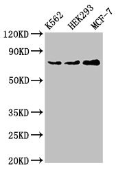
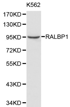
Western Blot Positive WB detected in: K562 whole cell lysate, HEK293 whole cell lysate, MCF-7 whole cell lysate All lanes: RALBP1 antibody at 4ug/ml Secondary Goat polyclonal to rabbit IgG at 1/50000 dilution Predicted band size: 77 kDa Observed band size: 77 kDa


")
Western Blot Positive WB detected in: K562 whole cell lysate, HEK293 whole cell lysate, MCF-7 whole cell lysate All lanes: RALBP1 antibody at 4ug/ml Secondary Goat polyclonal to rabbit IgG at 1/50000 dilution Predicted band size: 77 kDa Observed band size: 77 kDa
RALBP1 Antibody
CSB-PA618981LA01HU
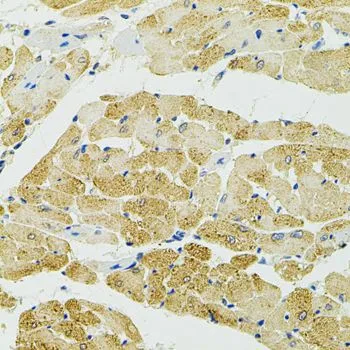
ApplicationsImmunoFluorescence, Western Blot, ELISA, ImmunoHistoChemistry
Product group Antibodies
ReactivityHuman
TargetRALBP1
Overview
- SupplierCusabio
- Product NameRALBP1 Antibody
- Delivery Days Customer20
- ApplicationsImmunoFluorescence, Western Blot, ELISA, ImmunoHistoChemistry
- CertificationResearch Use Only
- ClonalityPolyclonal
- ConjugateUnconjugated
- Gene ID10928
- Target nameRALBP1
- Target descriptionralA binding protein 1
- Target synonymsRIP1, RLIP1, RLIP76, ralA-binding protein 1, 76 kDa Ral-interacting protein, DNP-SG ATPase, dinitrophenyl S-glutathione ATPase, ral-interacting protein 1
- HostRabbit
- IsotypeIgG
- Protein IDQ15311
- Protein NameRalA-binding protein 1
- Scientific DescriptionCan activate specifically hydrolysis of GTP bound to RAC1 and CDC42, but not RALA. Mediates ATP-dependent transport of S-(2,4-dinitrophenyl)-glutathione (DNP-SG) and doxorubicin (DOX) and is the major ATP-dependent transporter of glutathione conjugates of electrophiles (GS-E) and DOX in erythrocytes. Can catalyze transport of glutathione conjugates and xenobiotics, and may contribute to the multidrug resistance phenomenon. Serves as a scaffold protein that brings together proteins forming an endocytotic complex during interphase and also with CDK1 to switch off endocytosis, One of its substrates would be EPN1/Epsin.
- ReactivityHuman
- Storage Instruction-20°C or -80°C
- UNSPSC41116161
Related products

Product group Antibodies
Anti-RALBP1 Antibody Picoband(r)A01403-1-CARRIER-FREE
ApplicationsFlow Cytometry, ImmunoFluorescence, Western Blot, ELISA, ImmunoCytoChemistry, ImmunoHistoChemistry
ReactivityHuman, Mouse, Rat
TargetRALBP1
- SizePrice
Product group Antibodies
Anti-RALBP1 Antibody144-01140
ApplicationsWestern Blot, ImmunoHistoChemistry
ReactivityHuman, Mouse, Rat
TargetRALBP1
- SizePrice
Product group Antibodies
Anti-RALBP1 AntibodyA29421
ApplicationsWestern Blot, ImmunoHistoChemistry
ReactivityHuman, Mouse, Rat
- SizePrice

Product group Antibodies
Anti-RALBP1 AntibodyHPA007622
ApplicationsImmunoCytoChemistry
ReactivityHuman
TargetRALBP1
- SizePrice
Product group Antibodies

RIP1 / RALBP1 AntibodyLS-C331290
ApplicationsWestern Blot, ImmunoHistoChemistry
ReactivityHuman, Mouse, Rat
TargetRALBP1
- SizePrice
Product group Antibodies
RALBP1 antibodyGTX55774
ApplicationsWestern Blot, ImmunoHistoChemistry, ImmunoHistoChemistry Paraffin
ReactivityHuman, Mouse, Rat
TargetRALBP1
- SizePrice
Product group Antibodies
RALBP1 Recombinant AntibodyBSM-61265R
ApplicationsFlow Cytometry, ImmunoFluorescence, Western Blot, ImmunoHistoChemistry, ImmunoHistoChemistry Frozen, ImmunoHistoChemistry Paraffin
TargetRALBP1
- SizePrice
Product group Antibodies
RALBP1 Polyclonal AntibodyCAC14895
ApplicationsImmunoFluorescence, Western Blot, ELISA, ImmunoHistoChemistry
TargetRALBP1
- SizePrice