


Immunohistochemical staining of human cerebral cortex shows strong cytoplasmic positivity in neurons.

Immunohistochemical staining of human cerebral cortex shows strong cytoplasmic positivity in neurons.
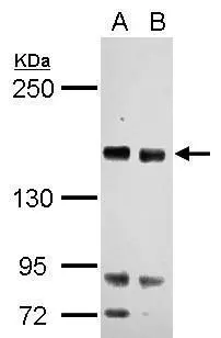
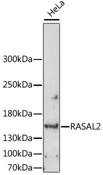
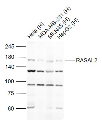
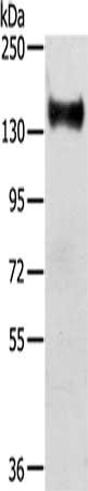
Anti-RASAL2 Antibody
HPA020453
ApplicationsWestern Blot, ImmunoHistoChemistry
Product group Antibodies
ReactivityHuman, Mouse
TargetRASAL2
Overview
- SupplierAtlas Antibodies
- Product NameAnti-RASAL2 Antibody
- Delivery Days Customer4
- ApplicationsWestern Blot, ImmunoHistoChemistry
- CertificationResearch Use Only
- ClonalityPolyclonal
- ConjugateUnconjugated
- Gene ID9462
- Target nameRASAL2
- Target descriptionRAS protein activator like 2
- Target synonymsNGAP, ras GTPase-activating protein nGAP, RASAL2/ACVR1 fusion, Ras protein activator like 1
- HostRabbit
- IsotypeIgG
- Protein IDQ9UJF2
- Protein NameRas GTPase-activating protein nGAP
- Scientific DescriptionRecombinant Protein Epitope Signature Tag (PrEST) antigen sequence
- ReactivityHuman, Mouse
- Storage Instruction-20°C,2°C to 8°C
- UNSPSC41116161
Datasheet
MSDS
Related products
Product group Antibodies
Anti-RASAL2 Antibody144-60923
ApplicationsWestern Blot
ReactivityHuman
TargetRASAL2
- SizePrice
Product group Antibodies
References
RASAL2 antibody [C2C3], C-termGTX120989
ApplicationsWestern Blot, ImmunoHistoChemistry, ImmunoHistoChemistry Paraffin
ReactivityHuman
TargetRASAL2
- SizePrice
Product group Antibodies
Anti-RASAL2 AntibodyA88417
ApplicationsImmunoFluorescence, Western Blot, ImmunoCytoChemistry
ReactivityHuman, Rat
- SizePrice
Product group Antibodies
RASAL2 Polyclonal AntibodyBS-21160R
ApplicationsWestern Blot, ELISA
ReactivityHuman, Mouse
TargetRASAL2
- SizePrice
Product group Antibodies

NGAP / RASAL2 AntibodyLS-C750092
ApplicationsWestern Blot
ReactivityHuman
TargetRASAL2
- SizePrice
Product group Antibodies
RASAL2 AntibodyCSB-PA151979
ApplicationsWestern Blot, ELISA
ReactivityHuman, Mouse
TargetRASAL2
- SizePrice

Product group Antibodies
Anti-RASAL2 Antibody Picoband(r)A07356-1-CARRIER-FREE
ApplicationsFlow Cytometry, Western Blot, ELISA
ReactivityHuman, Mouse, Rat
TargetRASAL2
- SizePrice