


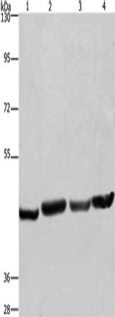
Figure 1. Western blot analysis of RBMY1A1 using anti-RBMY1A1 antibody (A06728-1). Electrophoresis was performed on a 5-20% SDS-PAGE gel at 70V (Stacking gel) / 90V (Resolving gel) for 2-3 hours. The sample well of each lane was loaded with 30 ug of sample under reducing conditions. Lane 1: human HEL whole cell lysates, Lane 2: rat brain tissue lysates, Lane 3: mouse testis tissue lysates, Lane 4: mouse brain tissue lysates, Lane 5: mouse NIH/3T3 whole cell lysates. After electrophoresis, proteins were transferred to a nitrocellulose membrane at 150 mA for 50-90 minutes. Blocked the membrane with 5% non-fat milk/TBS for 1.5 hour at RT. The membrane was incubated with rabbit anti-RBMY1A1 antigen affinity purified polyclonal antibody (Catalog # A06728-1) at 0.5 microg/mL overnight at 4°C, then washed with TBS-0.1%Tween 3 times with 5 minutes each and probed with a goat anti-rabbit IgG-HRP secondary antibody at a dilution of 1:5000 for 1.5 hour at RT. The signal is developed using an Enhanced Chemiluminescent detection (ECL) kit (Catalog # EK1002) with Tanon 5200 system. A specific band was detected for RBMY1A1 at approximately 51 kDa. The expected band size for RBMY1A1 is at 56 kDa.
 and anti-Tubulin beta antibody (M05613-4). RBMY1A1 and Tubulin beta was detected in an immunocytochemical section of Hela cells. Enzyme antigen retrieval was performed using IHC enzyme antigen retrieval reagent (AR0022) for 15 mins. The cells were blocked with 10% goat serum. And then incubated with 5 microg/mL rabbit anti-RBMY1A1 Antibody (A06728-1) and mouse anti-Tubulin beta Antibody (M05613-4) overnight at 4°C. Cy3 Conjugated Goat Anti-Rabbit IgG (BA1032) and DyLight®488 Conjugated Goat Anti-Mouse IgG (BA1126) were used as secondary antibody at 1:500 dilution and incubated for 30 minutes at 37°C. Visualize using a fluorescence microscope and filter sets appropriate for the label used.")
Figure 1. Western blot analysis of RBMY1A1 using anti-RBMY1A1 antibody (A06728-1). Electrophoresis was performed on a 5-20% SDS-PAGE gel at 70V (Stacking gel) / 90V (Resolving gel) for 2-3 hours. The sample well of each lane was loaded with 30 ug of sample under reducing conditions. Lane 1: human HEL whole cell lysates, Lane 2: rat brain tissue lysates, Lane 3: mouse testis tissue lysates, Lane 4: mouse brain tissue lysates, Lane 5: mouse NIH/3T3 whole cell lysates. After electrophoresis, proteins were transferred to a nitrocellulose membrane at 150 mA for 50-90 minutes. Blocked the membrane with 5% non-fat milk/TBS for 1.5 hour at RT. The membrane was incubated with rabbit anti-RBMY1A1 antigen affinity purified polyclonal antibody (Catalog # A06728-1) at 0.5 microg/mL overnight at 4°C, then washed with TBS-0.1%Tween 3 times with 5 minutes each and probed with a goat anti-rabbit IgG-HRP secondary antibody at a dilution of 1:5000 for 1.5 hour at RT. The signal is developed using an Enhanced Chemiluminescent detection (ECL) kit (Catalog # EK1002) with Tanon 5200 system. A specific band was detected for RBMY1A1 at approximately 51 kDa. The expected band size for RBMY1A1 is at 56 kDa.
Anti-RBMY1A1 Antibody Picoband(r)
A06728-1-CARRIER-FREE
ApplicationsImmunoFluorescence, Western Blot, ELISA, ImmunoCytoChemistry
Product group Antibodies
ReactivityHuman, Mouse, Rat
TargetRBMY1A1
Overview
- SupplierBoster Bio
- Product NameAnti-RBMY1A1 Antibody Picoband(r)
- Delivery Days Customer9
- ApplicationsImmunoFluorescence, Western Blot, ELISA, ImmunoCytoChemistry
- CertificationResearch Use Only
- ClonalityPolyclonal
- Concentration500 ug/ml
- Gene ID5940
- Target nameRBMY1A1
- Target descriptionRNA binding motif protein Y-linked family 1 member A1
- Target synonymsRBM, RBM1, RBM2, RBMY, RBMY1C, YRRM1, YRRM2, RNA-binding motif protein, Y chromosome, family 1 member A1, RNA-binding motif protein 2, RNA-binding motif protein, Y chromosome, family 1 member A1/C, Y chromosome RNA recognition motif 1, Y chromosome RNA recognition motif 2
- HostRabbit
- IsotypeIgG
- Protein IDP0DJD3
- Protein NameRNA-binding motif protein, Y chromosome, family 1 member A1
- Scientific DescriptionBoster Bio Anti-RBMY1A1 Antibody Picoband® catalog # A06728-1. Tested in ELISA, IF, ICC, WB applications. This antibody reacts with Human, Mouse, Rat. The brand Picoband indicates this is a premium antibody that guarantees superior quality, high affinity, and strong signals with minimal background in Western blot applications. Only our best-performing antibodies are designated as Picoband, ensuring unmatched performance.
- ReactivityHuman, Mouse, Rat
- Storage Instruction-20°C,2°C to 8°C
- UNSPSC12352203
Related products
Product group Antibodies
RBMY1A1 AntibodyCSB-PA289764
ApplicationsWestern Blot, ELISA
ReactivityHuman
TargetRBMY1A1
- SizePrice

Product group Antibodies
Anti-RBMY1A1 AntibodyA308659
ApplicationsImmunoFluorescence, ImmunoCytoChemistry, ImmunoHistoChemistry
ReactivityHuman, Mouse, Rat
- SizePrice
Product group Antibodies

RBMY1A1 Antibody (FITC)LS-C318086
ApplicationsWestern Blot, ELISA
ReactivityHuman
TargetRBMY1A1
- SizePrice
Product group Antibodies
Anti-RBMY1A1 AntibodyHPA001534
ApplicationsImmunoHistoChemistry
ReactivityHuman
TargetRBMY1A1
- SizePrice
Product group Antibodies
RBMY1A1 antibody [N1N3]GTX109967
ApplicationsWestern Blot
ReactivityHuman
TargetRBMY1A1
- SizePrice
Product group Antibodies
RBMY1A1 Polyclonal AntibodyCAC14154
ApplicationsImmunoFluorescence, Western Blot, ELISA, ImmunoHistoChemistry
ReactivityRat
TargetRBMY1A1
- SizePrice