


Figure 1. Western blot analysis of RHOB using anti-RHOB antibody (A01550-1). Electrophoresis was performed on a 5-20% SDS-PAGE gel at 70V (Stacking gel) / 90V (Resolving gel) for 2-3 hours. The sample well of each lane was loaded with 30 ug of sample under reducing conditions. Lane 1: human Hela whole cell lysates, Lane 2: rat brain tissue lysates, Lane 3: rat C6 whole cell lysates, Lane 4: mouse brain tissue lysates, Lane 5: monkey COS-7 whole cell lysates. After electrophoresis, proteins were transferred to a nitrocellulose membrane at 150 mA for 50-90 minutes. Blocked the membrane with 5% non-fat milk/TBS for 1.5 hour at RT. The membrane was incubated with rabbit anti-RHOB antigen affinity purified polyclonal antibody (Catalog # A01550-1) at 0.5 microg/mL overnight at 4°C, then washed with TBS-0.1%Tween 3 times with 5 minutes each and probed with a goat anti-rabbit IgG-HRP secondary antibody at a dilution of 1:5000 for 1.5 hour at RT. The signal is developed using an Enhanced Chemiluminescent detection (ECL) kit (Catalog # EK1002) with Tanon 5200 system. A specific band was detected for RHOB at approximately 22 kDa. The expected band size for RHOB is at 22 kDa.
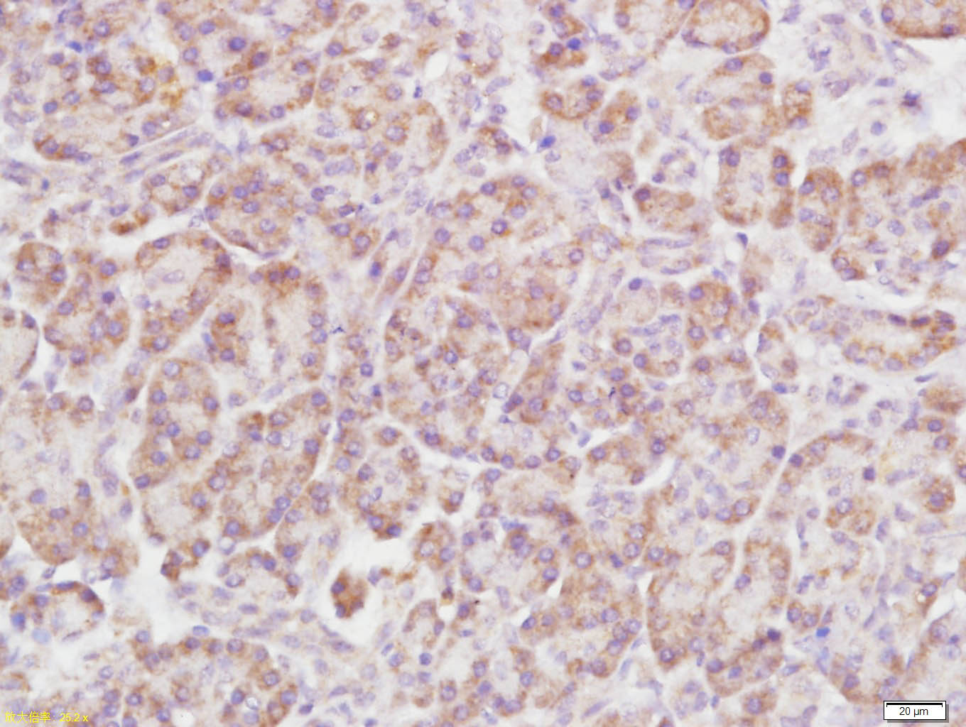
. RHOB was detected in paraffin-embedded section of human renal cancer tissue. Heat mediated antigen retrieval was performed in citrate buffer (pH6, epitope retrieval solution) for 20 mins. The tissue section was blocked with 10% goat serum. The tissue section was then incubated with 1microg/ml rabbit anti-RHOB Antibody (A01550-1) overnight at 4°C. Biotinylated goat anti-rabbit IgG was used as secondary antibody and incubated for 30 minutes at 37°C. The tissue section was developed using Strepavidin-Biotin-Complex (SABC)(Catalog # SA1022) with DAB as the chromogen.")
. RHOB was detected in paraffin-embedded section of human mammary cancer tissue. Heat mediated antigen retrieval was performed in citrate buffer (pH6, epitope retrieval solution) for 20 mins. The tissue section was blocked with 10% goat serum. The tissue section was then incubated with 1microg/ml rabbit anti-RHOB Antibody (A01550-1) overnight at 4°C. Biotinylated goat anti-rabbit IgG was used as secondary antibody and incubated for 30 minutes at 37°C. The tissue section was developed using Strepavidin-Biotin-Complex (SABC)(Catalog # SA1022) with DAB as the chromogen.")
. RHOB was detected in an immunocytochemical section of U20S cells. Enzyme antigen retrieval was performed using IHC enzyme antigen retrieval reagent (AR0022) for 15 mins. The cells were blocked with 10% goat serum. And then incubated with 5 microg/mL rabbit anti-RHOB Antibody (A01550-1) overnight at 4°C. DyLight®488 Conjugated Goat Anti-Rabbit IgG (BA1127) was used as secondary antibody at 1:100 dilution and incubated for 30 minutes at 37°C. The section was counterstained with DAPI. Visualize using a fluorescence microscope and filter sets appropriate for the label used.")
Figure 1. Western blot analysis of RHOB using anti-RHOB antibody (A01550-1). Electrophoresis was performed on a 5-20% SDS-PAGE gel at 70V (Stacking gel) / 90V (Resolving gel) for 2-3 hours. The sample well of each lane was loaded with 30 ug of sample under reducing conditions. Lane 1: human Hela whole cell lysates, Lane 2: rat brain tissue lysates, Lane 3: rat C6 whole cell lysates, Lane 4: mouse brain tissue lysates, Lane 5: monkey COS-7 whole cell lysates. After electrophoresis, proteins were transferred to a nitrocellulose membrane at 150 mA for 50-90 minutes. Blocked the membrane with 5% non-fat milk/TBS for 1.5 hour at RT. The membrane was incubated with rabbit anti-RHOB antigen affinity purified polyclonal antibody (Catalog # A01550-1) at 0.5 microg/mL overnight at 4°C, then washed with TBS-0.1%Tween 3 times with 5 minutes each and probed with a goat anti-rabbit IgG-HRP secondary antibody at a dilution of 1:5000 for 1.5 hour at RT. The signal is developed using an Enhanced Chemiluminescent detection (ECL) kit (Catalog # EK1002) with Tanon 5200 system. A specific band was detected for RHOB at approximately 22 kDa. The expected band size for RHOB is at 22 kDa.
Anti-RHOB Antibody
A01550-1
ApplicationsImmunoFluorescence, Western Blot, ImmunoCytoChemistry, ImmunoHistoChemistry
Product group Antibodies
ReactivityHuman, Monkey, Mouse, Rat
TargetRHOB
Overview
- SupplierBoster Bio
- Product NameAnti-RHOB Antibody
- Delivery Days Customer9
- ApplicationsImmunoFluorescence, Western Blot, ImmunoCytoChemistry, ImmunoHistoChemistry
- CertificationResearch Use Only
- ClonalityPolyclonal
- Concentration500 ug/ml
- Gene ID388
- Target nameRHOB
- Target descriptionras homolog family member B
- Target synonymsARH6, ARHB, MST081, MSTP081, RHOH6, rho-related GTP-binding protein RhoB, Aplysia RAS-related homolog 6, h6, oncogene RHO H6, ras homolog gene family, member B, rho cDNA clone 6
- HostRabbit
- IsotypeIgG
- Protein IDP62745
- Protein NameRho-related GTP-binding protein RhoB
- Scientific DescriptionBoster Bio Anti-RHOB Antibody Picoband® catalog # A01550-1. Tested in IF, IHC, ICC, WB applications. This antibody reacts with Human, Monkey, Mouse, Rat. The brand Picoband indicates this is a premium antibody that guarantees superior quality, high affinity, and strong signals with minimal background in Western blot applications. Only our best-performing antibodies are designated as Picoband, ensuring unmatched performance.
- ReactivityHuman, Monkey, Mouse, Rat
- Storage Instruction-20°C,2°C to 8°C
- UNSPSC12352203
Datasheet
MSDS
Related products
Product group Antibodies
ApplicationsImmunoPrecipitation, Western Blot, ImmunoCytoChemistry, ImmunoHistoChemistry
TargetRHOB
- SizePrice
Product group Antibodies
Rho B Polyclonal Antibodybs-11142R
ApplicationsImmunoFluorescence, ELISA, ImmunoCytoChemistry, ImmunoHistoChemistry, ImmunoHistoChemistry Frozen, ImmunoHistoChemistry Paraffin
ReactivityCanine, Chicken, Human, Mouse, Rabbit, Rat
TargetRHOB
- SizePrice
Product group Antibodies
Anti-RHOB Antibody144-02819
ApplicationsWestern Blot, ImmunoHistoChemistry
ReactivityHuman, Mouse
TargetRHOB
- SizePrice
Product group Antibodies
Anti-RHOB AntibodyA97935
ApplicationsWestern Blot, ELISA
ReactivityHuman, Mouse, Rat
- SizePrice
Product group Antibodies
RhoB antibodyGTX108600
ApplicationsWestern Blot, ImmunoHistoChemistry, ImmunoHistoChemistry Paraffin
ReactivityHuman, Mouse, Rat
TargetRHOB
- SizePrice
Product group Antibodies

RHOB AntibodyLS-C402119
ApplicationsWestern Blot, ELISA
ReactivityHuman, Mouse, Rat
TargetRHOB
- SizePrice
Product group Antibodies
RHOB AntibodyCSB-PA009684
ApplicationsWestern Blot, ELISA
ReactivityHuman, Mouse, Rat
TargetRHOB
- SizePrice
Product group Antibodies
Anti-RHOB Antibody Picoband(r)A01550-1-CARRIER-FREE
ApplicationsImmunoFluorescence, Western Blot, ImmunoCytoChemistry, ImmunoHistoChemistry
ReactivityHuman, Monkey, Mouse, Rat
TargetRHOB
- SizePrice