


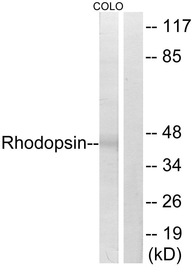
Western blot analysis of lysates from COLO cells, using Rhodopsin Antibody. The lane on the right is blocked with the synthesized peptide.

Western blot analysis of lysates from COLO cells, using Rhodopsin Antibody. The lane on the right is blocked with the synthesized peptide.
Anti-Rhodopsin Antibody
A30435
ApplicationsImmunoFluorescence, Western Blot, ELISA, ImmunoCytoChemistry, ImmunoHistoChemistry
Product group Antibodies
ReactivityHuman, Mouse, Rat
TargetRHO
Overview
- SupplierBoster Bio
- Product NameAnti-Rhodopsin Antibody
- Delivery Days Customer9
- ApplicationsImmunoFluorescence, Western Blot, ELISA, ImmunoCytoChemistry, ImmunoHistoChemistry
- CertificationResearch Use Only
- ClonalityPolyclonal
- Concentration0.5-1 mg/ml
- Gene ID6010
- Target nameRHO
- Target descriptionrhodopsin
- Target synonymsCSNBAD1, OPN2, RP4, rhodopsin, opsin 2, rod pigment, opsin-2
- HostRabbit
- IsotypeIgG
- Protein IDP08100
- Protein NameRhodopsin
- Scientific DescriptionBoster Bio Anti-Rhodopsin Antibody (Catalog# A30435). Tested in WB, IHC, ICC, IF, ELISA applications. This antibody reacts with Human, Mouse, Rat.
- ReactivityHuman, Mouse, Rat
- Storage Instruction-20°C,2°C to 8°C
- UNSPSC12352203
Related products
Product group Antibodies
Anti-Rhodopsin AntibodyA95174
ApplicationsWestern Blot, ELISA, ImmunoHistoChemistry
ReactivityHuman, Mouse, Rat
- SizePrice
Product group Antibodies

Anti-Rhodopsin [Rho 1D4]Ab00337-1.1
ApplicationsImmunoFluorescence, ImmunoPrecipitation, Western Blot, ELISA, ImmunoHistoChemistry, Labeling/Conjugation
ReactivityAmphibian, Bovine, Human, Mouse, Rat, Zebra Fish
TargetRHO
- SizePrice
Product group Antibodies
Anti-RHO Antibody144-63580
ApplicationsWestern Blot, ImmunoHistoChemistry
ReactivityHuman, Mouse, Rat
TargetRHO
- SizePrice

Product group Antibodies
ApplicationsWestern Blot, ImmunoHistoChemistry
ReactivityHuman, Rat
TargetRHO
- SizePrice
Product group Antibodies
Rhodopsin Recombinant Antibody, AbBy Fluor-350 ConjugatedBSM-61439R-BF350
ApplicationsImmunoFluorescence, Western Blot
ReactivityHuman, Rat
TargetRHO
- SizePrice
Product group Antibodies
RHO AntibodyCSB-PA003975
ApplicationsWestern Blot, ELISA
ReactivityHuman, Mouse, Rat
TargetRHO
- SizePrice
Product group Antibodies

Rhodopsin / RHO Antibody (C-Terminus)LS-C368503
ApplicationsWestern Blot
ReactivityHuman, Mouse
TargetRHO
- SizePrice
![Rhodopsin antibody detects Rhodopsin protein expression by immunohistochemical analysis. Sample: Frozen sectioned adult mouse retina. Green: Rhodopsin protein stained by Rhodopsin antibody (GTX129910) diluted at 1:250. Red: beta Tubulin 3/ TUJ1, stained by beta Tubulin 3/ TUJ1 antibody [GT11710] (GTX631836) diluted at 1:250. Blue: Fluoroshield with DAPI (GTX30920).](https://www.genetex.com/upload/website/prouct_img/normal/GTX129910/GTX129910_41927_20170214_IHC-Fr_w_23060523_396.webp)
Product group Antibodies
Rhodopsin antibodyGTX129910
ApplicationsWestern Blot, ImmunoHistoChemistry, ImmunoHistoChemistry Frozen, ImmunoHistoChemistry Paraffin
ReactivityHuman, Mouse, Rat
TargetRHO
- SizePrice