


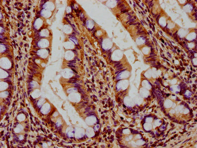
Immunohistochemical staining of human small intestine shows cytoplasmic positivity in glandular cells.

Immunohistochemical staining of human small intestine shows cytoplasmic positivity in glandular cells.
Anti-RHOT2 Antibody
HPA012895
ApplicationsImmunoCytoChemistry, ImmunoHistoChemistry
Product group Antibodies
ReactivityHuman
TargetRHOT2
Overview
- SupplierAtlas Antibodies
- Product NameAnti-RHOT2 Antibody
- Delivery Days Customer4
- ApplicationsImmunoCytoChemistry, ImmunoHistoChemistry
- CertificationResearch Use Only
- ClonalityPolyclonal
- ConjugateUnconjugated
- Gene ID89941
- Target nameRHOT2
- Target descriptionras homolog family member T2
- Target synonymsARHT2, C16orf39, MIRO-2, MIRO2, RASL, mitochondrial Rho GTPase 2, mitochondrial Rho (MIRO) GTPase 2, ras homolog gene family, member T2
- HostRabbit
- IsotypeIgG
- Protein IDQ8IXI1
- Protein NameMitochondrial Rho GTPase 2
- Scientific DescriptionRecombinant Protein Epitope Signature Tag (PrEST) antigen sequence
- ReactivityHuman
- Storage Instruction-20°C,2°C to 8°C
- UNSPSC41116161
Datasheet
MSDS
Related products

Product group Antibodies
Anti-MIRO2/RHOT2 Antibody Picoband(r)A08801-1-CARRIER-FREE
ApplicationsFlow Cytometry, ImmunoFluorescence, ImmunoPrecipitation, Western Blot, ELISA, ImmunoCytoChemistry, ImmunoHistoChemistry
ReactivityHuman, Mouse, Rat
TargetRHOT2
- SizePrice
Product group Antibodies
Anti-RHOT2 Antibody144-65555
ApplicationsWestern Blot
ReactivityHuman, Rat
TargetRHOT2
- SizePrice
Product group Antibodies
RHOT2 AntibodyCSB-PA810289LA01HU
ApplicationsELISA, ImmunoHistoChemistry
ReactivityHuman
TargetRHOT2
- SizePrice
Product group Antibodies

RHOT2 AntibodyLS-C408438
ApplicationsWestern Blot, ImmunoHistoChemistry
ReactivityHuman, Mouse, Rat
TargetRHOT2
- SizePrice

Product group Antibodies
Anti-RHOT2 AntibodyHPA012624
ApplicationsWestern Blot, ImmunoCytoChemistry, ImmunoHistoChemistry
ReactivityHuman
TargetRHOT2
- SizePrice
Product group Antibodies
Anti-RHOT2 AntibodyCAB2597
ApplicationsImmunoFluorescence, Western Blot, ELISA, ImmunoCytoChemistry, ImmunoHistoChemistry, ImmunoHistoChemistry Paraffin
ReactivityHuman
TargetRHOT2
- SizePrice