


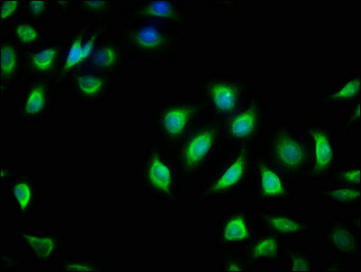
Immunohistochemical staining of human cerebellum shows moderate positivity in neuronal processes in molecular layer.
Immunohistochemical staining of human cerebellum shows moderate positivity in neuronal processes in molecular layer.
Anti-RIT2 Antibody
HPA072174
ApplicationsImmunoHistoChemistry
Product group Antibodies
ReactivityHuman
TargetRIT2
Overview
- SupplierAtlas Antibodies
- Product NameAnti-RIT2 Antibody
- Delivery Days Customer4
- ApplicationsImmunoHistoChemistry
- CertificationResearch Use Only
- ClonalityPolyclonal
- ConjugateUnconjugated
- Gene ID6014
- Target nameRIT2
- Target descriptionRas like without CAAX 2
- Target synonymsRIBA, RIN, ROC2, GTP-binding protein Rit2, GTP-binding protein Roc2, Ric-like, expressed in neurons, ras-like protein expressed in neurons, ras-like without CAAX protein 2
- HostRabbit
- IsotypeIgG
- Protein IDQ99578
- Protein NameGTP-binding protein Rit2
- Scientific DescriptionRecombinant Protein Epitope Signature Tag (PrEST) antigen sequence
- ReactivityHuman
- Storage Instruction-20°C,2°C to 8°C
- UNSPSC41116161
Datasheet
MSDS
Related products

Product group Antibodies
Anti-RIT2 Antibody Picoband(r)A07833-1-CARRIER-FREE
ApplicationsFlow Cytometry, Western Blot, ELISA
ReactivityHuman, Mouse, Rat
TargetRIT2
- SizePrice
Product group Antibodies
RIT2 AntibodyCSB-PA860769LA01HU
ApplicationsImmunoFluorescence, ELISA, ImmunoHistoChemistry
ReactivityHuman
TargetRIT2
- SizePrice
![WB analysis of various cell lines using GTX83710 RIT2 antibody [3F4]. Loading : 35 ug per lane](https://www.genetex.com/upload/website/prouct_img/normal/GTX83710/GTX83710_3838_WB_w_23061420_982.webp)
Product group Antibodies
RIT2 antibody [3F4]GTX83710
ApplicationsFlow Cytometry, Western Blot, ImmunoHistoChemistry, ImmunoHistoChemistry Paraffin
ReactivityCanine, Human, Monkey, Rat
TargetRIT2
- SizePrice
Product group Antibodies

RIT2 / RIN AntibodyLS-C748106
ApplicationsWestern Blot
ReactivityHuman, Mouse, Rat
TargetRIT2
- SizePrice