Bio-Connect
Immunofluorescent staining of human cell line A-431 shows localization to nucleoplasm.
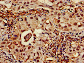
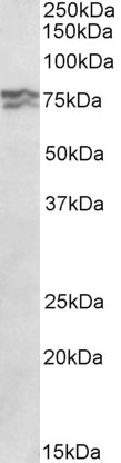
Anti-RPA1 Antibody

HPA046497
Atlas Antibodies
ApplicationsWestern Blot, ImmunoCytoChemistry
Product group Antibodies
ReactivityHuman
TargetRPA1
Sign in to order and to see your custom pricing.
Large volume orders?
Order with a bulk request

Overview

  • Supplier
    Atlas Antibodies
  • Product Name
    Anti-RPA1
  • Delivery Days Customer
    4
  • Applications
    Western Blot, ImmunoCytoChemistry
  • Certification
    Research Use Only
  • Clonality
    Polyclonal
  • Conjugate
    Unconjugated
  • Gene ID6117
  • Target name
    RPA1
  • Target description
    replication protein A1
  • Target synonyms
    HSSB; MST075; MSTP075; REPA1; replication factor A protein 1; replication protein A 70 kDa DNA-binding subunit; replication protein A1, 70kDa; RF-A; RF-A protein 1; RP-A; RP-A p70; RPA70; single-stranded DNA-binding protein
  • Host
    Rabbit
  • Isotype
    IgG
  • Protein IDP27694
  • Protein Name
    Replication protein A 70 kDa DNA-binding subunit
  • Scientific Description
    Recombinant Protein Epitope Signature Tag (PrEST) antigen sequence
  • Reactivity
    Human
  • Storage Instruction
    -20°C,2°C to 8°C
  • UNSPSC
    41116161

Related products

Product group Antibodies
RayBiotech
ApplicationsWestern Blot, ImmunoHistoChemistry
TargetRPA1
  • SizePrice
Product group Antibodies
Biomatik
ApplicationsWestern Blot, ELISA, ImmunoHistoChemistry
TargetRPA1
  • SizePrice
Product group Antibodies
Assay Genie
TargetRPA1
  • SizePrice
Immunohistochemistry of paraffin-embedded human lung cancer using CSB-PA020088LA01HU at dilution of 1:100
Product group Antibodies
Cusabio
RPA1 AntibodyCSB-PA020088LA01HU
ApplicationsWestern Blot, ELISA, ImmunoHistoChemistry
ReactivityHuman, Mouse, Rat
TargetRPA1
  • SizePrice
Product group Antibodies
Biosynth
  • SizePrice
Product group Antibodies
Bioss Antibodies
ApplicationsFlow Cytometry, ImmunoFluorescence, Western Blot, ImmunoCytoChemistry
TargetRPA1
  • SizePrice
Immunohistochemical staining of human placenta shows strong nuclear positivity in trophoblastic cells.
Product group Antibodies
Atlas Antibodies
ApplicationsImmunoHistoChemistry
ReactivityHuman
TargetRPA1
  • SizePrice
Immunohistochemical staining of human placenta shows strong nuclear positivity in trophoblastic cells.
Product group Antibodies
Atlas Antibodies
ApplicationsImmunoHistoChemistry
ReactivityHuman
TargetRPA1
  • SizePrice
Atlas Antibodies
Protein IDP27694
  • SizePrice
Figure 1. Western blot analysis of RPA70 using anti-RPA70 antibody (PB9886). Electrophoresis was performed on a 5-20% SDS-PAGE gel at 70V (Stacking gel) / 90V (Resolving gel) for 2-3 hours. The sample well of each lane was loaded with 30 ug of sample under reducing conditions. Lane 1: human Hela whole cell lysates, Lane 2: human K562 whole cell lysates, Lane 3: human HepG2 whole cell lysates, Lane 4: human 293T whole cell lysates. After electrophoresis, proteins were transferred to a nitrocellulose membrane at 150 mA for 50-90 minutes. Blocked the membrane with 5% non-fat milk/TBS for 1.5 hour at RT. The membrane was incubated with rabbit anti-RPA70 antigen affinity purified polyclonal antibody (Catalog # PB9886) at 0.5 microg/mL overnight at 4°C, then washed with TBS-0.1%Tween 3 times with 5 minutes each and probed with a goat anti-rabbit IgG-HRP secondary antibody at a dilution of 1:5000 for 1.5 hour at RT. The signal is developed using an Enhanced Chemiluminescent detection (ECL) kit (Catalog # EK1002) with Tanon 5200 system. A specific band was detected for RPA70 at approximately 70 kDa. The expected band size for RPA70 is at 68 kDa.
Product group Antibodies
Boster Bio
ApplicationsFlow Cytometry, ImmunoFluorescence, Western Blot, ImmunoCytoChemistry
TargetRPA1
  • SizePrice