

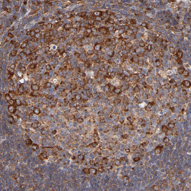
Immunohistochemical staining of human duodenum shows strong cytoplasmic positivity in glandular cells.
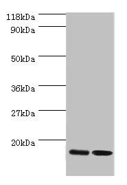
![Lane 1: Marker [kDa] 220, 112, 84, 47, 32, 26, 17. Lane 2: Human cell line RT-4. Lane 3: Human cell line U-251MG sp](https://atlasantibodies.s3.amazonaws.com/images/wb/hpa002649-wb-1.jpg "Lane 1: Marker [kDa] 220, 112, 84, 47, 32, 26, 17. Lane 2: Human cell line RT-4. Lane 3: Human cell line U-251MG sp")
Immunohistochemical staining of human duodenum shows strong cytoplasmic positivity in glandular cells.
Anti-RPL27 Antibody
HPA002649
ApplicationsWestern Blot, ImmunoHistoChemistry
Product group Antibodies
ReactivityHuman
TargetRPL27
Overview
- SupplierAtlas Antibodies
- Product NameAnti-RPL27 Antibody
- Delivery Days Customer4
- ApplicationsWestern Blot, ImmunoHistoChemistry
- CertificationResearch Use Only
- ClonalityPolyclonal
- ConjugateUnconjugated
- Gene ID6155
- Target nameRPL27
- Target descriptionribosomal protein L27
- Target synonymsDBA16, L27, eL27, large ribosomal subunit protein eL27, 60S ribosomal protein L27
- HostRabbit
- IsotypeIgG
- Protein IDP61353
- Protein NameLarge ribosomal subunit protein eL27
- Scientific DescriptionRecombinant Protein Epitope Signature Tag (PrEST) antigen sequence
- ReactivityHuman
- Storage Instruction-20°C,2°C to 8°C
- UNSPSC41116161
Datasheet
MSDS
Related products

Product group Antibodies
Anti-RPL27 Antibody Picoband(r)A09736-2-CARRIER-FREE
ApplicationsFlow Cytometry, ImmunoPrecipitation, Western Blot, ELISA
ReactivityHuman, Mouse, Rat
TargetRPL27
- SizePrice
Product group Antibodies
Anti-RPL27 Antibody144-60157
ApplicationsWestern Blot
ReactivityHuman, Mouse
TargetRPL27
- SizePrice
Product group Antibodies

RPL27 / Ribosomal Protein L27 AntibodyLS-C748104
ApplicationsWestern Blot
ReactivityHuman, Mouse
TargetRPL27
- SizePrice
Product group Antibodies
Rpl27 Polyclonal AntibodyCAC08902
ApplicationsImmunoFluorescence, Western Blot, ELISA, ImmunoHistoChemistry
TargetRPL27
- SizePrice
Product group Antibodies
RPL27 AntibodyCSB-PA02345A0RB
ApplicationsImmunoFluorescence, Western Blot, ELISA, ImmunoHistoChemistry
ReactivityHuman
TargetRPL27
- SizePrice
Product group Antibodies
RPL27 antibodyGTX66153
ApplicationsImmunoFluorescence, Western Blot, ImmunoCytoChemistry
ReactivityHuman, Mouse, Rat
TargetRPL27
- SizePrice
Product group Antibodies
Anti-RPL27 AntibodyCAB13044
ApplicationsImmunoFluorescence, Western Blot, ELISA, ImmunoCytoChemistry
ReactivityHuman
TargetRPL27
- SizePrice