


Immunohistochemical staining of human cervix, uterine shows strong cytoplasmic positivity in glandular cells.

Immunohistochemical staining of human cervix, uterine shows strong cytoplasmic positivity in glandular cells.
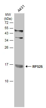
Anti-RPS25 Antibody
HPA031801
ApplicationsWestern Blot, ImmunoCytoChemistry, ImmunoHistoChemistry
Product group Antibodies
ReactivityHuman, Mouse, Rat
TargetRPS25
Overview
- SupplierAtlas Antibodies
- Product NameAnti-RPS25 Antibody
- Delivery Days Customer4
- ApplicationsWestern Blot, ImmunoCytoChemistry, ImmunoHistoChemistry
- CertificationResearch Use Only
- ClonalityPolyclonal
- ConjugateUnconjugated
- Gene ID6230
- Target nameRPS25
- Target descriptionribosomal protein S25
- Target synonymsS25, eS25, small ribosomal subunit protein eS25, 40S ribosomal protein S25
- HostRabbit
- IsotypeIgG
- Protein IDP62851
- Protein NameSmall ribosomal subunit protein eS25
- Scientific DescriptionRecombinant Protein Epitope Signature Tag (PrEST) antigen sequence
- ReactivityHuman, Mouse, Rat
- Storage Instruction-20°C,2°C to 8°C
- UNSPSC41116161
Datasheet
MSDS
Related products
Product group Antibodies
RPS25 AntibodyCSB-PA00644A0RB
ApplicationsELISA
ReactivityHuman
TargetRPS25
- SizePrice

Product group Antibodies
Anti-RPS25 Antibody Picoband(r)A09150-3-CARRIER-FREE
ApplicationsFlow Cytometry, Western Blot, ELISA, ImmunoHistoChemistry
ReactivityHuman, Mouse, Rat
TargetRPS25
- SizePrice
Product group Antibodies
Anti-RPS25 Antibody144-60993
ApplicationsWestern Blot
ReactivityHuman, Mouse, Rat
TargetRPS25
- SizePrice

Product group Antibodies
Anti-RPS25 AntibodyA96995
ApplicationsELISA, ImmunoHistoChemistry
ReactivityHuman, Mouse, Rat
- SizePrice
Product group Antibodies

RPS25 / Ribosomal Protein S25 AntibodyLS-C770368
ApplicationsWestern Blot
ReactivityHuman
TargetRPS25
- SizePrice

Product group Antibodies
Anti-RPS25 AntibodyHPA078683
ApplicationsImmunoCytoChemistry
ReactivityHuman
TargetRPS25
- SizePrice
Product group Antibodies
RPS25 antibodyGTX101526
ApplicationsWestern Blot
ReactivityHuman
TargetRPS25
- SizePrice