

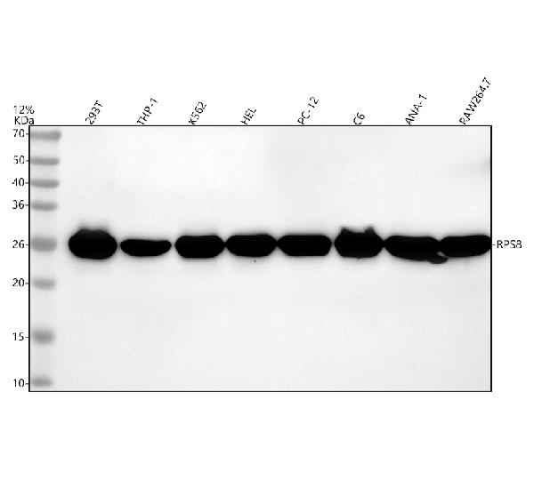
Figure 1. Western blot analysis of RPS8 using anti-RPS8 antibody (M07839). Electrophoresis was performed on a 5-20% SDS-PAGE gel at 70V (Stacking gel) / 90V (Resolving gel) for 2-3 hours. The sample well of each lane was loaded with 30 ug of sample under reducing conditions. Lane 1: human 293T whole cell lysates, Lane 2: human THP-1 whole cell lysates, Lane 3: human K562 whole cell lysates, Lane 4: human HEL whole cell lysates, Lane 5: rat PC-12 whole cell lysates, Lane 6: rat C6 whole cell lysates, Lane 7: mouse ANA-1 whole cell lysates, Lane 8: mouse RAW264.7 whole cell lysates. After electrophoresis, proteins were transferred to a nitrocellulose membrane at 150 mA for 50-90 minutes. Blocked the membrane with 5% non-fat milk/TBS for 1.5 hour at RT. The membrane was incubated with rabbit anti-RPS8 antigen affinity purified monoclonal antibody (M07839) at 1:500 overnight at 4°C, then washed with TBS-0.1%Tween 3 times with 5 minutes each and probed with a goat anti-rabbit IgG-HRP secondary antibody at a dilution of 1:500 for 1.5 hour at RT. The signal is developed using an Enhanced Chemiluminescent detection (ECL) kit (Catalog # EK1002) with Tanon 5200 system. A specific band was detected for RPS8 at approximately 26 kDa. The expected band size for RPS8 is at 24 kDa.
 analysis using the Antibody at 1:50 dilution. (wb at 1:1K dilution)")
Figure 1. Western blot analysis of RPS8 using anti-RPS8 antibody (M07839). Electrophoresis was performed on a 5-20% SDS-PAGE gel at 70V (Stacking gel) / 90V (Resolving gel) for 2-3 hours. The sample well of each lane was loaded with 30 ug of sample under reducing conditions. Lane 1: human 293T whole cell lysates, Lane 2: human THP-1 whole cell lysates, Lane 3: human K562 whole cell lysates, Lane 4: human HEL whole cell lysates, Lane 5: rat PC-12 whole cell lysates, Lane 6: rat C6 whole cell lysates, Lane 7: mouse ANA-1 whole cell lysates, Lane 8: mouse RAW264.7 whole cell lysates. After electrophoresis, proteins were transferred to a nitrocellulose membrane at 150 mA for 50-90 minutes. Blocked the membrane with 5% non-fat milk/TBS for 1.5 hour at RT. The membrane was incubated with rabbit anti-RPS8 antigen affinity purified monoclonal antibody (M07839) at 1:500 overnight at 4°C, then washed with TBS-0.1%Tween 3 times with 5 minutes each and probed with a goat anti-rabbit IgG-HRP secondary antibody at a dilution of 1:500 for 1.5 hour at RT. The signal is developed using an Enhanced Chemiluminescent detection (ECL) kit (Catalog # EK1002) with Tanon 5200 system. A specific band was detected for RPS8 at approximately 26 kDa. The expected band size for RPS8 is at 24 kDa.
Anti-RPS8 Rabbit Monoclonal Antibody
M07839
ApplicationsFlow Cytometry, ImmunoFluorescence, ImmunoPrecipitation, Western Blot, ImmunoCytoChemistry
Product group Antibodies
ReactivityHuman, Mouse, Rat
TargetRPS8
Overview
- SupplierBoster Bio
- Product NameAnti-RPS8 Rabbit Monoclonal Antibody
- Delivery Days Customer9
- ApplicationsFlow Cytometry, ImmunoFluorescence, ImmunoPrecipitation, Western Blot, ImmunoCytoChemistry
- CertificationResearch Use Only
- ClonalityMonoclonal
- Clone ID30R72
- Gene ID6202
- Target nameRPS8
- Target descriptionribosomal protein S8
- Target synonymsS8, eS8, small ribosomal subunit protein eS8, 40S ribosomal protein S8, OK/SW-cl.83
- HostRabbit
- IsotypeIgG
- Protein IDP62241
- Protein NameSmall ribosomal subunit protein eS8
- Scientific DescriptionBoster Bio Anti-RPS8 Rabbit Monoclonal Antibody catalog # M07839. Tested in WB, ICC/IF, IP, Flow Cytometry applications. This antibody reacts with Human, Mouse, Rat.
- ReactivityHuman, Mouse, Rat
- Storage Instruction-20°C
- UNSPSC12352203
Related products
Product group Antibodies
Anti-RPS8 AntibodyA97921
ApplicationsWestern Blot, ELISA
ReactivityHuman, Mouse, Rat
- SizePrice
Product group Antibodies
Anti-RPS8 (N-term) Antibody102-27608
ApplicationsFlow Cytometry, Western Blot
TargetRPS8
- SizePrice
Product group Antibodies
RPS8 Recombinant AntibodyBSM-62765R
ApplicationsFlow Cytometry, ImmunoFluorescence, ImmunoPrecipitation, Western Blot, ImmunoCytoChemistry
ReactivityHuman, Mouse, Rat
TargetRPS8
- SizePrice
Product group Antibodies
RPS8 AntibodyCSB-PA004007
ApplicationsWestern Blot, ELISA, ImmunoHistoChemistry
ReactivityHuman, Mouse, Rat
TargetRPS8
- SizePrice
Product group Antibodies
Rps8 Polyclonal AntibodyCAC08814
ApplicationsWestern Blot, ELISA
ReactivityMouse
TargetRPS8
- SizePrice
Product group Antibodies
RPS8 antibody [N1C3]GTX120300
ApplicationsWestern Blot
ReactivityHuman, Zebra Fish
TargetRPS8
- SizePrice
Product group Antibodies

RPS8 / Ribosomal Protein S8 AntibodyLS-C396473
ApplicationsELISA
ReactivityHuman
TargetRPS8
- SizePrice
Product group Antibodies
Anti-RPS8 AntibodyCAB18377
ApplicationsImmunoFluorescence, Western Blot, ELISA, ImmunoCytoChemistry
ReactivityHuman
TargetRPS8
- SizePrice