

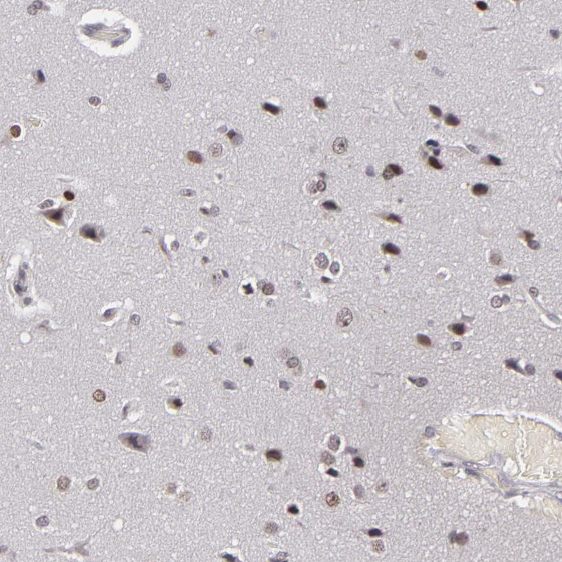
Immunohistochemical staining of human cerebral cortex shows strong nuclear positivity in neurons.

Immunohistochemical staining of human cerebral cortex shows strong nuclear positivity in neurons.
Anti-RRP7A Antibody
HPA001586
ApplicationsImmunoCytoChemistry, ImmunoHistoChemistry
Product group Antibodies
ReactivityHuman
TargetRRP7A
Overview
- SupplierAtlas Antibodies
- Product NameAnti-RRP7A Antibody
- Delivery Days Customer4
- ApplicationsImmunoCytoChemistry, ImmunoHistoChemistry
- CertificationResearch Use Only
- ClonalityPolyclonal
- ConjugateUnconjugated
- Gene ID27341
- Target nameRRP7A
- Target descriptionribosomal RNA processing 7 homolog A
- Target synonymsBK126B4.3, CGI-96, MCPH28, Rrp7, ribosomal RNA-processing protein 7 homolog A, CTA-126B4.5, gastric cancer antigen Zg14
- HostRabbit
- IsotypeIgG
- Protein IDQ9Y3A4
- Protein NameRibosomal RNA-processing protein 7 homolog A
- Scientific DescriptionRecombinant Protein Epitope Signature Tag (PrEST) antigen sequence
- ReactivityHuman
- Storage Instruction-20°C,2°C to 8°C
- UNSPSC41116161
Datasheet
MSDS
Related products

Product group Antibodies
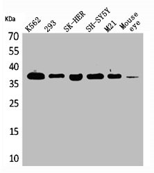
Anti-RRP7A Antibody Picoband(r)A15583-2-CARRIER-FREE
ApplicationsFlow Cytometry, ImmunoFluorescence, Western Blot, ELISA, ImmunoCytoChemistry, ImmunoHistoChemistry
ReactivityHuman, Monkey
TargetRRP7A
- SizePrice
Product group Antibodies
Anti-RRP7A (C-term) Antibody102-21321
ApplicationsWestern Blot
TargetRRP7A
- SizePrice
Product group Antibodies
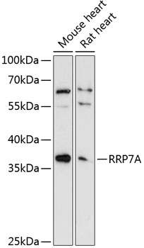
Anti-RRP7A AntibodyA89242
ApplicationsWestern Blot
ReactivityMouse, Rat
- SizePrice
Product group Antibodies
RRP7A AntibodyCSB-PA004778
ApplicationsWestern Blot, ELISA
ReactivityHuman, Mouse, Rat
TargetRRP7A
- SizePrice
Product group Antibodies

CGI-96 / RRP7A AntibodyLS-C411151
ApplicationsImmunoFluorescence, Western Blot, ImmunoHistoChemistry
ReactivityHuman, Mouse, Rat
TargetRRP7A
- SizePrice

Product group Antibodies
Anti-RRP7A AntibodyHPA046768
ApplicationsImmunoCytoChemistry
ReactivityHuman
TargetRRP7A
- SizePrice
Product group Antibodies
CTA-126B4.3 antibody, N-termGTX47308
ApplicationsWestern Blot
ReactivityHuman
TargetRRP7A
- SizePrice