

Anti-S100A9 [IID4B10]
Ab02468-1.1
ApplicationsWestern Blot, ELISA, ImmunoHistoChemistry
Product group Antibodies
ReactivityHuman
TargetS100A9
Overview
- SupplierAbsolute Antibody
- Product NameAnti-S100A9 [IID4B10]
- Delivery Days Customer7
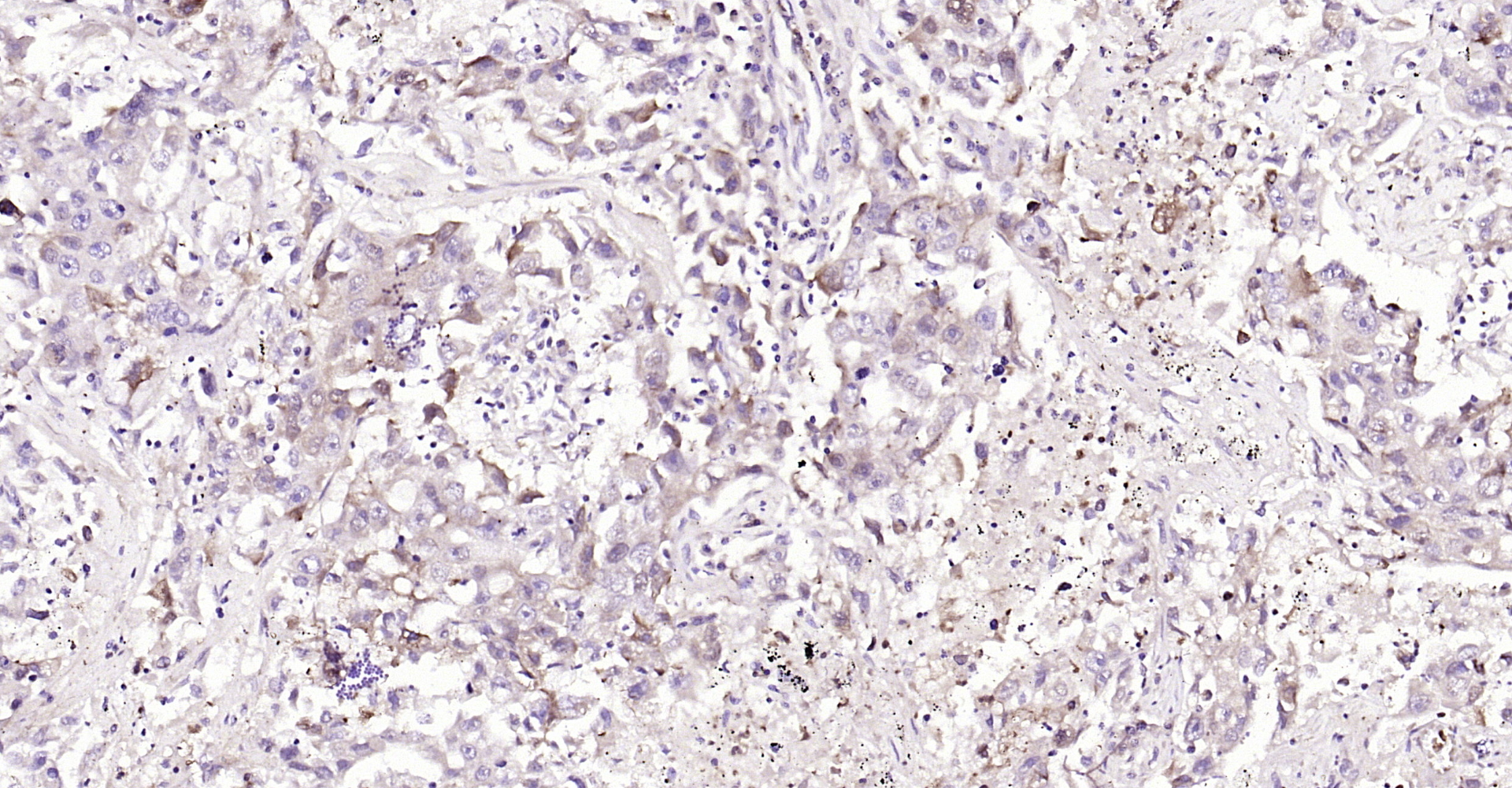
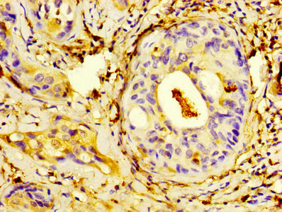
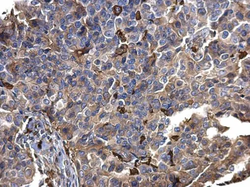
- Application Supplier NoteThis antibody was used for ELISA on recombinant S100A9 protein (CN104894074). Western blot was performed on S100A9, S100A8, S100A12 and S100A13 using this antibody (CN104894074). Immunohistochemistry was performed on human breast cancer, colon cancer, and hepatocellular carcinoma tissues using this antibody (CN104894074).
- ApplicationsWestern Blot, ELISA, ImmunoHistoChemistry
- Applications SupplierELISA; IHC; WB
- CertificationResearch Use Only
- ClonalityMonoclonal
- Clone IDIID4B10
- Gene ID6280
- Target nameS100A9
- Target descriptionS100 calcium binding protein A9
- Target synonyms60B8AG, CAGB, CFAG, CGLB, L1AG, LIAG, MAC387, MIF, MRP14, NIF, P14, S100-A9, protein S100-A9, MRP-14, calgranulin B, calprotectin L1H subunit, leukocyte L1 complex heavy chain, migration inhibitory factor-related protein 14
- HostMouse
- IsotypeIgG1
- Protein IDP06702
- Protein NameProtein S100-A9
- ReactivityHuman
- Reactivity SupplierHuman
- Reactivity Supplier NoteThe original antibody was raised by immunizing Balb/c mice with human blood cells.
- Storage Instruction-20°C,2°C to 8°C
- UNSPSC41116161
Related products
Product group Antibodies
ApplicationsWestern Blot, ELISA, ImmunoHistoChemistry
- SizePrice
Product group Antibodies
Anti-S100A9 Antibody130-10511
ApplicationsELISA
ReactivityHuman
TargetS100A9
- SizePrice

Product group Antibodies
Anti-S100A9 AntibodyAMAB91690
ApplicationsImmunoHistoChemistry
ReactivityHuman
TargetS100A9
- SizePrice
Product group Antibodies
S100A9 Polyclonal AntibodyCAC07212
ApplicationsImmunoFluorescence, ELISA
TargetS100A9
- SizePrice
Product group Antibodies
S100-A9 Polyclonal AntibodyBS-2697R
ApplicationsImmunoFluorescence, ELISA, ImmunoCytoChemistry, ImmunoHistoChemistry, ImmunoHistoChemistry Frozen, ImmunoHistoChemistry Paraffin
ReactivityHuman, Mouse, Rat
TargetS100A9
- SizePrice
Product group Antibodies
S100A9 AntibodyCSB-PA020642HA01HU
ApplicationsImmunoFluorescence, ELISA, ImmunoHistoChemistry
ReactivityHuman
TargetS100A9
- SizePrice
Product group Antibodies
S100A9 antibodyGTX129575
ApplicationsImmunoFluorescence, Western Blot, ImmunoCytoChemistry, ImmunoHistoChemistry, ImmunoHistoChemistry Paraffin
ReactivityHuman
TargetS100A9
- SizePrice
Product group Antibodies

Goat anti-S100A9EB09146
ApplicationsFlow Cytometry, ImmunoFluorescence, Western Blot, ELISA, ImmunoHistoChemistry
ReactivityHuman
TargetS100A9
- SizePrice