


Immunofluorescent staining of human cell line RT4 shows localization to nucleoplasm.
Immunofluorescent staining of human cell line RT4 shows localization to nucleoplasm.
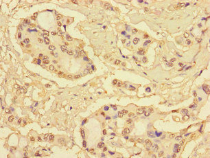
Anti-S100P Antibody
HPA075251
ApplicationsImmunoCytoChemistry
Product group Antibodies
ReactivityHuman
TargetS100P
Overview
- SupplierAtlas Antibodies
- Product NameAnti-S100P Antibody
- Delivery Days Customer4
- ApplicationsImmunoCytoChemistry
- CertificationResearch Use Only
- ClonalityPolyclonal
- ConjugateUnconjugated
- Gene ID6286
- Target nameS100P
- Target descriptionS100 calcium binding protein P
- Target synonymsMIG9, protein S100-P, migration-inducing gene 9 protein, protein S100-E
- HostRabbit
- IsotypeIgG
- Protein IDP25815
- Protein NameProtein S100-P
- Scientific DescriptionRecombinant Protein Epitope Signature Tag (PrEST) antigen sequence
- ReactivityHuman
- Storage Instruction-20°C,2°C to 8°C
- UNSPSC41116161
Datasheet
MSDS
Related products
Product group Antibodies
S100P AntibodyCSB-PA020645LA01HU
ApplicationsImmunoFluorescence, ELISA, ImmunoHistoChemistry
ReactivityHuman
TargetS100P
- SizePrice

Product group Antibodies
Anti-S100P Antibody Picoband(r)A01963-2-CARRIER-FREE
ApplicationsFlow Cytometry, ImmunoFluorescence, Western Blot, ImmunoCytoChemistry
ReactivityHuman
TargetS100P
- SizePrice
Product group Antibodies
Anti-S100P Antibody130-10036
ApplicationsELISA
ReactivityHuman
TargetS100P
- SizePrice
Product group Antibodies
Anti-S100P AntibodyA99598
ApplicationsWestern Blot, ELISA, ImmunoHistoChemistry
ReactivityHuman
- SizePrice
Product group Antibodies

S100P Antibody (Preservative Free)LS-C149199
ApplicationsELISA
ReactivityHuman
TargetS100P
- SizePrice

Product group Antibodies
Anti-S100P AntibodyHPA019502
ApplicationsImmunoHistoChemistry
ReactivityHuman
TargetS100P
- SizePrice
Product group Antibodies
Anti-S100P AntibodyHPA019502
ApplicationsImmunoHistoChemistry
ReactivityHuman
TargetS100P
- SizePrice