


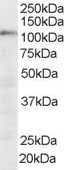
Figure 1. Western blot analysis of SART1 using anti-SART1 antibody (A05414-1). Electrophoresis was performed on a 5-20% SDS-PAGE gel at 70V (Stacking gel) / 90V (Resolving gel) for 2-3 hours. The sample well of each lane was loaded with 30 ug of sample under reducing conditions. Lane 1: human Hela whole cell lysates, Lane 2: human MCF-7 whole cell lysates, Lane 3: human MOLT-4 whole cell lysates, Lane 4: rat testis tissue lysates, Lane 5: mouse testis tissue lysates, Lane 6: mouse thymus tissue lysates. After electrophoresis, proteins were transferred to a nitrocellulose membrane at 150 mA for 50-90 minutes. Blocked the membrane with 5% non-fat milk/TBS for 1.5 hour at RT. The membrane was incubated with rabbit anti-SART1 antigen affinity purified polyclonal antibody (Catalog # A05414-1) at 0.5 microg/mL overnight at 4°C, then washed with TBS-0.1%Tween 3 times with 5 minutes each and probed with a goat anti-rabbit IgG-HRP secondary antibody at a dilution of 1:5000 for 1.5 hour at RT. The signal is developed using an Enhanced Chemiluminescent detection (ECL) kit (Catalog # EK1002) with Tanon 5200 system. A specific band was detected for SART1 at approximately 110 kDa. The expected band size for SART1 is at 90 kDa.
. SART1 was detected in a paraffin-embedded section of human colorectal adenocarcinoma tissue. Heat mediated antigen retrieval was performed in EDTA buffer (pH 8.0, epitope retrieval solution). The tissue section was blocked with 10% goat serum. The tissue section was then incubated with 2 microg/ml rabbit anti-SART1 Antibody (A05414-1) overnight at 4°C. Peroxidase Conjugated Goat Anti-rabbit IgG was used as secondary antibody and incubated for 30 minutes at 37°C. The tissue section was developed using HRP Conjugated Rabbit IgG Super Vision Assay Kit (Catalog # SV0002) with DAB as the chromogen.")
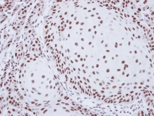
. SART1 was detected in a paraffin-embedded section of human ovarian cancer tissue. Heat mediated antigen retrieval was performed in EDTA buffer (pH 8.0, epitope retrieval solution). The tissue section was blocked with 10% goat serum. The tissue section was then incubated with 2 microg/ml rabbit anti-SART1 Antibody (A05414-1) overnight at 4°C. Peroxidase Conjugated Goat Anti-rabbit IgG was used as secondary antibody and incubated for 30 minutes at 37°C. The tissue section was developed using HRP Conjugated Rabbit IgG Super Vision Assay Kit (Catalog # SV0002) with DAB as the chromogen.")
. SART1 was detected in a paraffin-embedded section of human breast cancer tissue. Heat mediated antigen retrieval was performed in EDTA buffer (pH 8.0, epitope retrieval solution). The tissue section was blocked with 10% goat serum. The tissue section was then incubated with 2 microg/ml rabbit anti-SART1 Antibody (A05414-1) overnight at 4°C. Peroxidase Conjugated Goat Anti-rabbit IgG was used as secondary antibody and incubated for 30 minutes at 37°C. The tissue section was developed using HRP Conjugated Rabbit IgG Super Vision Assay Kit (Catalog # SV0002) with DAB as the chromogen.")
. SART1 was detected in a paraffin-embedded section of human laryngeal squamous cell carcinoma tissue. Heat mediated antigen retrieval was performed in EDTA buffer (pH 8.0, epitope retrieval solution). The tissue section was blocked with 10% goat serum. The tissue section was then incubated with 2 microg/ml rabbit anti-SART1 Antibody (A05414-1) overnight at 4°C. Peroxidase Conjugated Goat Anti-rabbit IgG was used as secondary antibody and incubated for 30 minutes at 37°C. The tissue section was developed using HRP Conjugated Rabbit IgG Super Vision Assay Kit (Catalog # SV0002) with DAB as the chromogen.")
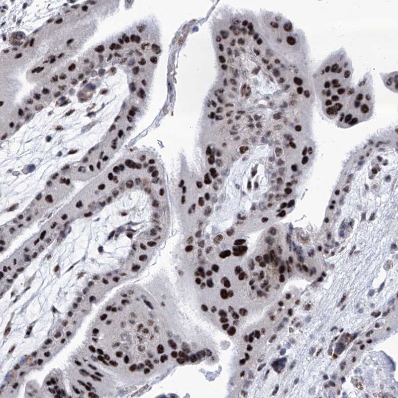
. SART1 was detected in a paraffin-embedded section of human lung cancer tissue. Heat mediated antigen retrieval was performed in EDTA buffer (pH 8.0, epitope retrieval solution). The tissue section was blocked with 10% goat serum. The tissue section was then incubated with 2 microg/ml rabbit anti-SART1 Antibody (A05414-1) overnight at 4°C. Peroxidase Conjugated Goat Anti-rabbit IgG was used as secondary antibody and incubated for 30 minutes at 37°C. The tissue section was developed using HRP Conjugated Rabbit IgG Super Vision Assay Kit (Catalog # SV0002) with DAB as the chromogen.")
 and anti-Beta Tubulin antibody (M01857-3). SART1 was detected in immunocytochemical section of A549 cell. Enzyme antigen retrieval was performed using IHC enzyme antigen retrieval reagent (AR0022) for 15 mins. The cells were blocked with 10% goat serum. And then incubated with 5 microg/mL rabbit anti-SART1 Antibody (A05414-1) and mouse anti-Beta Tubulin antibody (M01857-3) overnight at 4°C. Cy3 Conjugated Goat Anti-Rabbit IgG (BA1032) and DyLight®488 Conjugated Goat Anti-Mouse IgG (BA1126) were used as secondary antibody at 1:100 dilution and incubated for 30 minutes at 37°C. The section was counterstained with DAPI. Visualize using a fluorescence microscope and filter sets appropriate for the label used.")
. Overlay histogram showing U20S cells stained with A05414-1 (Blue line). To facilitate intracellular staining, cells were fixed with 4% paraformaldehyde and permeabilized with permeabilization buffer. The cells were blocked with 10% normal goat serum. And then incubated with rabbit anti-SART1 Antibody (A05414-1, 1 microg/1x106 cells) for 30 min at 20°C. DyLight®488 conjugated goat anti-rabbit IgG (BA1127, 5-10 microg/1x106 cells) was used as secondary antibody for 30 minutes at 20°C. Isotype control antibody (Green line) was rabbit IgG (1 microg/1x106) used under the same conditions. Unlabelled sample without incubation with primary antibody and secondary antibody (Red line) was used as a blank control.")
. SART1 was detected in a paraffin-embedded section of human breast cancer tissue. Heat mediated antigen retrieval was performed in EDTA buffer (pH 8.0, epitope retrieval solution). The tissue section was blocked with 10% goat serum. The tissue section was then incubated with 5 microg/mL rabbit anti-SART1 Antibody (A05414-1) overnight at 4°C. DyLight®550 Conjugated Goat Anti-Rabbit IgG (BA1135) was used as secondary antibody at 1:500 dilution and incubated for 30 minutes at 37°C. The section was counterstained with DAPI. Visualize using a fluorescence microscope and filter sets appropriate for the label used.")
Figure 1. Western blot analysis of SART1 using anti-SART1 antibody (A05414-1). Electrophoresis was performed on a 5-20% SDS-PAGE gel at 70V (Stacking gel) / 90V (Resolving gel) for 2-3 hours. The sample well of each lane was loaded with 30 ug of sample under reducing conditions. Lane 1: human Hela whole cell lysates, Lane 2: human MCF-7 whole cell lysates, Lane 3: human MOLT-4 whole cell lysates, Lane 4: rat testis tissue lysates, Lane 5: mouse testis tissue lysates, Lane 6: mouse thymus tissue lysates. After electrophoresis, proteins were transferred to a nitrocellulose membrane at 150 mA for 50-90 minutes. Blocked the membrane with 5% non-fat milk/TBS for 1.5 hour at RT. The membrane was incubated with rabbit anti-SART1 antigen affinity purified polyclonal antibody (Catalog # A05414-1) at 0.5 microg/mL overnight at 4°C, then washed with TBS-0.1%Tween 3 times with 5 minutes each and probed with a goat anti-rabbit IgG-HRP secondary antibody at a dilution of 1:5000 for 1.5 hour at RT. The signal is developed using an Enhanced Chemiluminescent detection (ECL) kit (Catalog # EK1002) with Tanon 5200 system. A specific band was detected for SART1 at approximately 110 kDa. The expected band size for SART1 is at 90 kDa.
Anti-SART1 Antibody Picoband(r)
A05414-1-CARRIER-FREE
ApplicationsFlow Cytometry, ImmunoFluorescence, Western Blot, ELISA, ImmunoCytoChemistry, ImmunoHistoChemistry
Product group Antibodies
ReactivityHuman, Mouse, Rat
TargetSART1
Overview
- SupplierBoster Bio
- Product NameAnti-SART1 Antibody Picoband(r)
- Delivery Days Customer9
- ApplicationsFlow Cytometry, ImmunoFluorescence, Western Blot, ELISA, ImmunoCytoChemistry, ImmunoHistoChemistry
- CertificationResearch Use Only
- ClonalityPolyclonal
- Concentration500 ug/ml
- Gene ID9092
- Target nameSART1
- Target descriptionspliceosome associated factor 1, recruiter of U4/U6.U5 tri-snRNP
- Target synonymsAra1, HAF, HOMS1, SART1259, SNRNP110, Snu66, U4/U6.U5 tri-snRNP-associated protein 1, IgE autoantigen, SART-1, SART1(259) protein, SART1(800) protein, SART1, U4/U6.U5 tri-snRNP-associated protein 1, SNU66 homolog, U4/U6.U5 tri-snRNP-associated 110 kDa protein, hSART-1, hSnu66, hypoxia associated factor, small nuclear ribonucleoprotein 110kDa (U4/U6.U5), squamous cell carcinoma antigen recognised by T cells, squamous cell carcinoma antigen recognized by T-cells 1
- HostRabbit
- IsotypeIgG
- Protein IDO43290
- Protein NameU4/U6.U5 tri-snRNP-associated protein 1
- Scientific DescriptionBoster Bio Anti-SART1 Antibody Picoband® catalog # A05414-1. Tested in ELISA, Flow Cytometry, IF, IHC, ICC, WB applications. This antibody reacts with Human, Mouse, Rat. The brand Picoband indicates this is a premium antibody that guarantees superior quality, high affinity, and strong signals with minimal background in Western blot applications. Only our best-performing antibodies are designated as Picoband, ensuring unmatched performance.
- ReactivityHuman, Mouse, Rat
- Storage Instruction-20°C,2°C to 8°C
- UNSPSC12352203
Related products
Product group Antibodies
Anti-SART1 AntibodyA83261
ApplicationsWestern Blot, ELISA
ReactivityHuman
- SizePrice
Product group Antibodies
Anti-SART1 Antibody144-08569
ApplicationsImmunoPrecipitation, Western Blot
ReactivityHuman, Mouse, Rat
TargetSART1
- SizePrice
Product group Antibodies
SART1 Polyclonal AntibodyBS-7874R
ApplicationsImmunoFluorescence, Western Blot, ELISA, ImmunoCytoChemistry, ImmunoHistoChemistry, ImmunoHistoChemistry Frozen, ImmunoHistoChemistry Paraffin
ReactivityBovine, Human, Mouse, Porcine, Rat, Sheep
TargetSART1
- SizePrice
Product group Antibodies

Goat anti-SART1EB05742
ApplicationsWestern Blot, ELISA
ReactivityBovine, Human, Mouse, Rat
TargetSART1
- SizePrice
Product group Antibodies

SART1 AntibodyLS-C410104
ApplicationsImmunoPrecipitation, Western Blot
ReactivityHuman, Mouse, Rat
TargetSART1
- SizePrice
Product group Antibodies
SART1 antibody [N2C1], InternalGTX103371
ApplicationsImmunoFluorescence, Western Blot, ImmunoCytoChemistry, ImmunoHistoChemistry, ImmunoHistoChemistry Paraffin
ReactivityHuman, Mouse
TargetSART1
- SizePrice
Product group Antibodies
Anti-SART1 AntibodyHPA031188
ApplicationsWestern Blot, ImmunoCytoChemistry, ImmunoHistoChemistry
ReactivityHuman
TargetSART1
- SizePrice
Product group Antibodies
Anti-SART1 AntibodyCAB8569
ApplicationsImmunoPrecipitation, Western Blot, ELISA, ImmunoHistoChemistry, ImmunoHistoChemistry Paraffin
ReactivityHuman
TargetSART1
- SizePrice