Bio-Connect
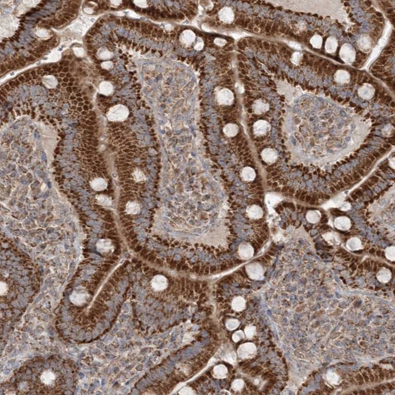
Immunohistochemical staining of human colon shows strong cytoplasmic positivity in endothelial cells.

Anti-SCLT1 Antibody

HPA036560
Atlas Antibodies
ApplicationsImmunoHistoChemistry
Product group Antibodies
ReactivityHuman
TargetSCLT1
Sign in to order and to see your custom pricing.
Large volume orders?
Order with a bulk request

Overview

  • Supplier
    Atlas Antibodies
  • Product Name
    Anti-SCLT1
  • Delivery Days Customer
    4
  • Applications
    ImmunoHistoChemistry
  • Certification
    Research Use Only
  • Clonality
    Polyclonal
  • Conjugate
    Unconjugated
  • Gene ID132320
  • Target name
    SCLT1
  • Target description
    sodium channel and clathrin linker 1
  • Target synonyms
    CAP1A; CAP-1A; sodium channel and clathrin linker 1; sodium channel-associated protein 1
  • Host
    Rabbit
  • Isotype
    IgG
  • Protein IDQ96NL6
  • Protein Name
    Sodium channel and clathrin linker 1
  • Scientific Description
    Recombinant Protein Epitope Signature Tag (PrEST) antigen sequence
  • Reactivity
    Human
  • Storage Instruction
    -20°C,2°C to 8°C
  • UNSPSC
    41116161

Related products

Immunohistochemistry of paraffin-embedded human liver tissue using CSB-PA020826LA01HU at dilution of 1:100
Product group Antibodies
Cusabio
SCLT1 AntibodyCSB-PA020826LA01HU
ApplicationsELISA, ImmunoHistoChemistry
ReactivityHuman
TargetSCLT1
  • SizePrice
Product group Antibodies
LifeSpan BioSciences
SCLT1 AntibodyLS-C396453
ApplicationsELISA, ImmunoHistoChemistry, ImmunoHistoChemistry Paraffin
TargetSCLT1
  • SizePrice
Immunohistochemical staining of human duodenum shows strong cytoplasmic positivity, with a granular pattern in glandular cells.
Product group Antibodies
Atlas Antibodies
ApplicationsWestern Blot, ImmunoHistoChemistry
ReactivityHuman
TargetSCLT1
  • SizePrice
Figure 1. Western blot analysis of SCLT1 using anti-SCLT1 antibody (A11417-1). Electrophoresis was performed on a 5-20% SDS-PAGE gel at 70V (Stacking gel) / 90V (Resolving gel) for 2-3 hours. The sample well of each lane was loaded with 30 ug of sample under reducing conditions. Lane 1: human 293T whole cell lysates, Lane 2: human COLO320 whole cell lysates. After electrophoresis, proteins were transferred to a nitrocellulose membrane at 150 mA for 50-90 minutes. Blocked the membrane with 5% non-fat milk/TBS for 1.5 hour at RT. The membrane was incubated with rabbit anti-SCLT1 antigen affinity purified polyclonal antibody (Catalog # A11417-1) at 0.5 microg/mL overnight at 4°C, then washed with TBS-0.1%Tween 3 times with 5 minutes each and probed with a goat anti-rabbit IgG-HRP secondary antibody at a dilution of 1:5000 for 1.5 hour at RT. The signal is developed using an Enhanced Chemiluminescent detection (ECL) kit (Catalog # EK1002) with Tanon 5200 system. A specific band was detected for SCLT1 at approximately 75 kDa. The expected band size for SCLT1 is at 81 kDa.
Product group Antibodies
Boster Bio
Anti-SCLT1 Antibody Picoband(r)A11417-1-CARRIER-FREE
ApplicationsFlow Cytometry, Western Blot, ELISA
TargetSCLT1
  • SizePrice
Atlas Antibodies
Protein IDQ96NL6
  • SizePrice
Atlas Antibodies
Protein IDQ96NL6
  • SizePrice