Bio-Connect
Immunofluorescent staining of human cell line HeLa shows localization to nucleus.
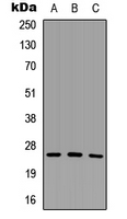
Anti-SCNM1 Antibody

HPA054324
Atlas Antibodies
ApplicationsImmunoCytoChemistry
Product group Antibodies
TargetSCNM1
Sign in to order and to see your custom pricing.
Large volume orders?
Order with a bulk request

Overview

  • Supplier
    Atlas Antibodies
  • Product Name
    Anti-SCNM1
  • Delivery Days Customer
    4
  • Applications
    ImmunoCytoChemistry
  • Certification
    Research Use Only
  • Clonality
    Polyclonal
  • Conjugate
    Unconjugated
  • Gene ID79005
  • Target name
    SCNM1
  • Target description
    sodium channel modifier 1
  • Target synonyms
    sodium channel modifier 1
  • Host
    Rabbit
  • Isotype
    IgG
  • Protein IDQ9BWG6
  • Protein Name
    Sodium channel modifier 1
  • Scientific Description
    Recombinant Protein Epitope Signature Tag (PrEST) antigen sequence
  • Storage Instruction
    -20°C,2°C to 8°C
  • UNSPSC
    12352203

Related products

Immunohistochemistry of paraffin-embedded human breast cancer using CSB-PA020847LA01HU at dilution of 1:100
Product group Antibodies
Cusabio
SCNM1 AntibodyCSB-PA020847LA01HU
ApplicationsELISA, ImmunoHistoChemistry
ReactivityHuman
TargetSCNM1
  • SizePrice
Atlas Antibodies
Protein IDQ9BWG6
  • SizePrice
Atlas Antibodies
Protein IDQ9BWG6
  • SizePrice
Immunohistochemical staining of human uterus shows strong cytoplasmic positivity.
Product group Antibodies
Atlas Antibodies
ApplicationsImmunoHistoChemistry
TargetSCNM1
  • SizePrice
Immunohistochemical staining of human uterus shows strong cytoplasmic positivity.
Product group Antibodies
Atlas Antibodies
ApplicationsImmunoHistoChemistry
TargetSCNM1
  • SizePrice
Product group Antibodies
LifeSpan BioSciences
SCNM1 AntibodyLS-C376545
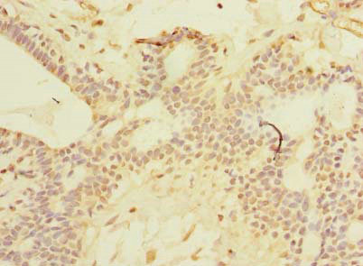
ApplicationsELISA, ImmunoHistoChemistry, ImmunoHistoChemistry Paraffin
TargetSCNM1
  • SizePrice
Figure 1. Western blot analysis of SCNM1 using anti-SCNM1 antibody (A10999-1). Electrophoresis was performed on a 5-20% SDS-PAGE gel at 70V (Stacking gel) / 90V (Resolving gel) for 2-3 hours. The sample well of each lane was loaded with 30 ug of sample under reducing conditions. Lane 1: human THP-1 whole cell lysates, Lane 2: rat lung tissue lysates, Lane 3: rat stomach tissue lysates, Lane 4: mouse lung tissue lysates, Lane 5: mouse stomach tissue lysates. After electrophoresis, proteins were transferred to a nitrocellulose membrane at 150 mA for 50-90 minutes. Blocked the membrane with 5% non-fat milk/TBS for 1.5 hour at RT. The membrane was incubated with rabbit anti-SCNM1 antigen affinity purified polyclonal antibody (Catalog # A10999-1) at 0.5 microg/mL overnight at 4°C, then washed with TBS-0.1%Tween 3 times with 5 minutes each and probed with a goat anti-rabbit IgG-HRP secondary antibody at a dilution of 1:5000 for 1.5 hour at RT. The signal is developed using an Enhanced Chemiluminescent detection (ECL) kit (Catalog # EK1002) with Tanon 5200 system. A specific band was detected for SCNM1 at approximately 26 kDa. The expected band size for SCNM1 is at 26 kDa.
Product group Antibodies
Boster Bio
Anti-SCNM1 Antibody Picoband(r)A10999-1-CARRIER-FREE
ApplicationsFlow Cytometry, Western Blot, ELISA, ImmunoHistoChemistry
TargetSCNM1
  • SizePrice
WB analysis of HeLa (A), Raw264.7 (B), H9C2 (C) whole cell lysates using GTX56073 SCNM1 antibody.
Product group Antibodies
GeneTex
ApplicationsWestern Blot
TargetSCNM1
  • SizePrice