

anti-SDHB antibody
ARG40979
ApplicationsImmunoFluorescence, Western Blot, ImmunoCytoChemistry, ImmunoHistoChemistry, ImmunoHistoChemistry Paraffin
Product group Antibodies
ReactivityHuman
TargetSDHB
Overview
- SupplierArigo Biolaboratories
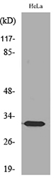
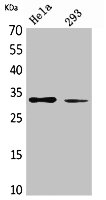
- Product Nameanti-SDHB antibody
- Delivery Days Customer23
- Application Supplier NoteIHC-P: Antigen Retrieval: Heat mediation was performed in Citrate buffer (pH 6.0, epitope retrieval solution) for 20 min.* The dilutions indicate recommended starting dilutions and the optimal dilutions or concentrations should be determined by the scientist.
- ApplicationsImmunoFluorescence, Western Blot, ImmunoCytoChemistry, ImmunoHistoChemistry, ImmunoHistoChemistry Paraffin
- CertificationResearch Use Only
- ClonalityPolyclonal
- Concentration0.5 mg/ml
- ConjugateUnconjugated
- Gene ID6390
- Target nameSDHB
- Target descriptionsuccinate dehydrogenase complex iron sulfur subunit B
- Target synonymsCWS2, IP, MC2DN4, PGL4, PPGL4, SDH, SDH1, SDH2, SDHIP, succinate dehydrogenase [ubiquinone] iron-sulfur subunit, mitochondrial, iron-sulfur subunit of complex II, malate dehydrogenase [quinone] iron-sulfur subunit, succinate dehydrogenase complex, subunit B, iron sulfur (Ip)
- HostRabbit
- IsotypeIgG
- Protein IDP21912
- Protein NameSuccinate dehydrogenase [ubiquinone] iron-sulfur subunit, mitochondrial
- Scientific DescriptionComplex II of the respiratory chain, which is specifically involved in the oxidation of succinate, carries electrons from FADH to CoQ. The complex is composed of four nuclear-encoded subunits and is localized in the mitochondrial inner membrane. The iron-sulfur subunit is highly conserved and contains three cysteine-rich clusters which may comprise the iron-sulfur centers of the enzyme. Sporadic and familial mutations in this gene result in paragangliomas and pheochromocytoma, and support a link between mitochondrial dysfunction and tumorigenesis. [provided by RefSeq, Jul 2008]
- ReactivityHuman
- Storage Instruction-20°C
- UNSPSC41116161
Related products
Product group Antibodies
Anti-SDHB AntibodyA97916
ApplicationsWestern Blot, ELISA
ReactivityHuman, Mouse, Rat
- SizePrice

Product group Antibodies
Anti-SDHB Antibody Picoband(r)A01090-CARRIER-FREE
ApplicationsImmunoFluorescence, Western Blot, ELISA, ImmunoCytoChemistry, ImmunoHistoChemistry
ReactivityHuman, Mouse, Rat
TargetSDHB
- SizePrice
Product group Antibodies
Anti-SDHB Antibody144-10821
ApplicationsWestern Blot, ImmunoHistoChemistry
ReactivityHuman, Mouse, Rat
TargetSDHB
- SizePrice

Product group Antibodies
Anti-SDHB AntibodyAMAB90705
ApplicationsWestern Blot, ImmunoHistoChemistry
ReactivityHuman
TargetSDHB
- SizePrice

Product group Antibodies
SDHB Recombinant AntibodyBSM-54180R
ApplicationsFlow Cytometry, ImmunoFluorescence, Western Blot, ImmunoCytoChemistry, ImmunoHistoChemistry, ImmunoHistoChemistry Frozen, ImmunoHistoChemistry Paraffin
ReactivityHuman, Mouse, Rat
TargetSDHB
- SizePrice
Product group Antibodies

Goat anti-SDHB, BiotinylatedEB06868-B
ApplicationsWestern Blot, ELISA
ReactivityHuman
TargetSDHB
- SizePrice
Product group Antibodies
Sdhb Recombinant AntibodyCAC12569
ApplicationsFlow Cytometry, Western Blot, ELISA, ImmunoHistoChemistry
ReactivityMouse, Rat
TargetSDHB
- SizePrice
Product group Antibodies
SDHB AntibodyCSB-PA005802
ApplicationsWestern Blot, ELISA
ReactivityHuman, Mouse, Rat
TargetSDHB
- SizePrice
Product group Antibodies

SDHB AntibodyLS-C406761
ApplicationsWestern Blot, ELISA, ImmunoHistoChemistry
ReactivityHuman, Mouse, Rat
TargetSDHB
- SizePrice
![SDHB antibody [C2C3], C-term detects SDHB protein at mitochondria by immunofluorescent analysis. Sample: HeLa cells were fixed in 4% paraformaldehyde at RT for 15 min. Green: SDHB protein stained by SDHB antibody [C2C3], C-term (GTX104628) diluted at 1:500. Blue: Hoechst 33342 staining.](https://www.genetex.com/upload/website/prouct_img/normal/GTX104628/GTX104628_41752_20160602_IFA_w_23060120_212.webp)
Product group Antibodies
SDHB antibody [C2C3], C-termGTX104628
ApplicationsImmunoFluorescence, Western Blot, ImmunoCytoChemistry, ImmunoHistoChemistry, ImmunoHistoChemistry Paraffin
ReactivityHuman, Mouse
TargetSDHB
- SizePrice