


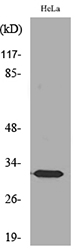
Figure 1. Western blot analysis of SDHB using anti-SDHB antibody (M01090-2). Electrophoresis was performed on a 5-20% SDS-PAGE gel at 70V (Stacking gel) / 90V (Resolving gel) for 2-3 hours. The sample well of each lane was loaded with 50ug of sample under reducing conditions. Lane 1: human HEK293 whole cell lysates, Lane 2: human HepG2 whole cell lysates, Lane 3: human THP-1 whole cell lysates, After Electrophoresis, proteins were transferred to a Nitrocellulose membrane at 150mA for 50-90 minutes. Blocked the membrane with 5% Non-fat Milk/ TBS for 1.5 hour at RT. The membrane was incubated with mouse anti-SDHB antigen affinity purified monoclonal antibody (Catalog # M01090-2) at 0.5 microg/mL overnight at 4°C, then washed with TBS-0.1%Tween 3 times with 5 minutes each and probed with a goat anti-mouse IgG-HRP secondary antibody at a dilution of 1:10000 for 1.5 hour at RT. The signal is developed using an Enhanced Chemiluminescent detection (ECL) kit (Catalog # EK1001) with Tanon 5200 system. A specific band was detected for SDHB at approximately 29KD. The expected band size for SDHB is at 29KD.
. SDHB was detected in paraffin-embedded section of human intestinal cancer tissues. Heat mediated antigen retrieval was performed in citrate buffer (pH6, epitope retrieval solution) for 20 mins. The tissue section was blocked with 10% goat serum. The tissue section was then incubated with 1microg/ml mouse anti-SDHB Antibody (M01090-2) overnight at 4°C. Biotinylated goat anti-mouse IgG was used as secondary antibody and incubated for 30 minutes at 37°C. The tissue section was developed using Strepavidin-Biotin-Complex (SABC)(Catalog # SA1021) with DAB as the chromogen.")
. SDHB was detected in paraffin-embedded section of human mammary cancer tissues. Heat mediated antigen retrieval was performed in citrate buffer (pH6, epitope retrieval solution) for 20 mins. The tissue section was blocked with 10% goat serum. The tissue section was then incubated with 1microg/ml mouse anti-SDHB Antibody (M01090-2) overnight at 4°C. Biotinylated goat anti-mouse IgG was used as secondary antibody and incubated for 30 minutes at 37°C. The tissue section was developed using Strepavidin-Biotin-Complex (SABC)(Catalog # SA1021) with DAB as the chromogen.")
. SDHB was detected in paraffin-embedded section of mouse cardiac muscle tissues. Heat mediated antigen retrieval was performed in citrate buffer (pH6, epitope retrieval solution) for 20 mins. The tissue section was blocked with 10% goat serum. The tissue section was then incubated with 1microg/ml mouse anti-SDHB Antibody (M01090-2) overnight at 4°C. Biotinylated goat anti-mouse IgG was used as secondary antibody and incubated for 30 minutes at 37°C. The tissue section was developed using Strepavidin-Biotin-Complex (SABC)(Catalog # SA1021) with DAB as the chromogen.")
. SDHB was detected in paraffin-embedded section of rat cardiac muscle tissues. Heat mediated antigen retrieval was performed in citrate buffer (pH6, epitope retrieval solution) for 20 mins. The tissue section was blocked with 10% goat serum. The tissue section was then incubated with 1microg/ml mouse anti-SDHB Antibody (M01090-2) overnight at 4°C. Biotinylated goat anti-mouse IgG was used as secondary antibody and incubated for 30 minutes at 37°C. The tissue section was developed using Strepavidin-Biotin-Complex (SABC)(Catalog # SA1021) with DAB as the chromogen.")
Figure 1. Western blot analysis of SDHB using anti-SDHB antibody (M01090-2). Electrophoresis was performed on a 5-20% SDS-PAGE gel at 70V (Stacking gel) / 90V (Resolving gel) for 2-3 hours. The sample well of each lane was loaded with 50ug of sample under reducing conditions. Lane 1: human HEK293 whole cell lysates, Lane 2: human HepG2 whole cell lysates, Lane 3: human THP-1 whole cell lysates, After Electrophoresis, proteins were transferred to a Nitrocellulose membrane at 150mA for 50-90 minutes. Blocked the membrane with 5% Non-fat Milk/ TBS for 1.5 hour at RT. The membrane was incubated with mouse anti-SDHB antigen affinity purified monoclonal antibody (Catalog # M01090-2) at 0.5 microg/mL overnight at 4°C, then washed with TBS-0.1%Tween 3 times with 5 minutes each and probed with a goat anti-mouse IgG-HRP secondary antibody at a dilution of 1:10000 for 1.5 hour at RT. The signal is developed using an Enhanced Chemiluminescent detection (ECL) kit (Catalog # EK1001) with Tanon 5200 system. A specific band was detected for SDHB at approximately 29KD. The expected band size for SDHB is at 29KD.
Anti-SDHB Antibody Picoband(r) (monoclonal, 11I3)
M01090-2-FITC
ApplicationsWestern Blot, ImmunoHistoChemistry
Product group Antibodies
ReactivityHuman, Mouse, Rat
TargetSDHB
Overview
- SupplierBoster Bio
- Product NameAnti-SDHB Antibody Picoband(r) (monoclonal, 11I3)
- Delivery Days Customer9
- ApplicationsWestern Blot, ImmunoHistoChemistry
- CertificationResearch Use Only
- ClonalityMonoclonal
- Clone ID11I3
- ConjugateFITC
- Gene ID6390
- Target nameSDHB
- Target descriptionsuccinate dehydrogenase complex iron sulfur subunit B
- Target synonymsCWS2, IP, MC2DN4, PGL4, PPGL4, SDH, SDH1, SDH2, SDHIP, succinate dehydrogenase [ubiquinone] iron-sulfur subunit, mitochondrial, iron-sulfur subunit of complex II, malate dehydrogenase [quinone] iron-sulfur subunit, succinate dehydrogenase complex, subunit B, iron sulfur (Ip)
- HostMouse
- IsotypeIgG1
- Protein IDP21912
- Protein NameSuccinate dehydrogenase [ubiquinone] iron-sulfur subunit, mitochondrial
- Scientific DescriptionBoster Bio Anti-SDHB Antibody Picoband® (monoclonal, 11I3) catalog # M01090-2. Tested in IHC, WB applications. This antibody reacts with Human, Mouse, Rat. The brand Picoband indicates this is a premium antibody that guarantees superior quality, high affinity, and strong signals with minimal background in Western blot applications. Only our best-performing antibodies are designated as Picoband, ensuring unmatched performance.
- ReactivityHuman, Mouse, Rat
- Storage Instruction-20°C,2°C to 8°C
- UNSPSC12352203
Related products

Product group Antibodies
SDHB Recombinant AntibodyBSM-54180R
ApplicationsImmunoFluorescence, Western Blot, ImmunoHistoChemistry, ImmunoHistoChemistry Frozen, ImmunoHistoChemistry Paraffin
ReactivityHuman, Mouse, Rat
TargetSDHB
- SizePrice
Product group Antibodies
Sdhb Recombinant AntibodyCAC12569
ApplicationsFlow Cytometry, Western Blot, ELISA, ImmunoHistoChemistry
ReactivityMouse, Rat
TargetSDHB
- SizePrice

Product group Antibodies
Anti-SDHB AntibodyAMAB90705
ApplicationsWestern Blot, ImmunoHistoChemistry
ReactivityHuman
TargetSDHB
- SizePrice
Product group Antibodies
Anti-SDHB Antibody144-10821
ApplicationsWestern Blot, ImmunoHistoChemistry
ReactivityHuman, Mouse, Rat
TargetSDHB
- SizePrice
Product group Antibodies
Anti-SDHB AntibodyA97916
ApplicationsWestern Blot, ELISA
ReactivityHuman, Mouse, Rat
- SizePrice
Product group Antibodies

ApplicationsWestern Blot, ELISA
ReactivityHuman
TargetSDHB
- SizePrice
Product group Antibodies
References
SDHB antibody [C2C3], C-termGTX104628
ApplicationsImmunoFluorescence, Western Blot, ImmunoCytoChemistry, ImmunoHistoChemistry, ImmunoHistoChemistry Paraffin
ReactivityHuman, Mouse
TargetSDHB
- SizePrice
Product group Antibodies

SDHB AntibodyLS-C406761
ApplicationsWestern Blot, ELISA, ImmunoHistoChemistry
ReactivityHuman, Mouse, Rat
TargetSDHB
- SizePrice
Product group Antibodies
SDHB AntibodyCSB-PA005802
ApplicationsWestern Blot, ELISA
ReactivityHuman, Mouse, Rat
TargetSDHB
- SizePrice