


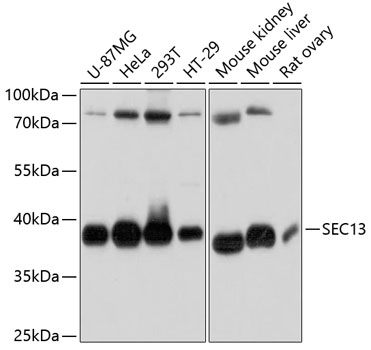
Figure 1. Western blot analysis of SEC13L1/SEC13 using anti-SEC13L1/SEC13 antibody (A04346-1). Electrophoresis was performed on a 5-20% SDS-PAGE gel at 70V (Stacking gel) / 90V (Resolving gel) for 2-3 hours. The sample well of each lane was loaded with 30 ug of sample under reducing conditions. Lane 1: human MCF-7 whole cell lysates, Lane 2: human MDA-MB-453 whole cell lysates, Lane 3: human HepG2 whole cell lysates, Lane 4: human K562 whole cell lysates, Lane 5: rat brain tissue lysates, Lane 6: rat small intestine tissue lysates, Lane 7: mosue brain tissue lysates, Lane 8: mouse small intestine tissue lysates. After electrophoresis, proteins were transferred to a nitrocellulose membrane at 150 mA for 50-90 minutes. Blocked the membrane with 5% non-fat milk/TBS for 1.5 hour at RT. The membrane was incubated with rabbit anti-SEC13L1/SEC13 antigen affinity purified polyclonal antibody (Catalog # A04346-1) at 0.5 microg/mL overnight at 4°C, then washed with TBS-0.1%Tween 3 times with 5 minutes each and probed with a goat anti-rabbit IgG-HRP secondary antibody at a dilution of 1:5000 for 1.5 hour at RT. The signal is developed using an Enhanced Chemiluminescent detection (ECL) kit (Catalog # EK1002) with Tanon 5200 system. A specific band was detected for SEC13L1/SEC13 at approximately 36 kDa. The expected band size for SEC13L1/SEC13 is at 36 kDa.
. SEC13L1/SEC13 was detected in an immunocytochemical section of A549 cells. Enzyme antigen retrieval was performed using IHC enzyme antigen retrieval reagent (AR0022) for 15 mins. The cells were blocked with 10% goat serum. And then incubated with 5 microg/mL rabbit anti-SEC13L1/SEC13 Antibody (A04346-1) overnight at 4°C. DyLight?488 Conjugated Goat Anti-Rabbit IgG (BA1127) was used as secondary antibody at 1:100 dilution and incubated for 30 minutes at 37°C. The section was counterstained with DAPI. Visualize using a fluorescence microscope and filter sets appropriate for the label used.")
. Overlay histogram showing HL-60 cells stained with A04346-1 (Blue line). To facilitate intracellular staining, cells were fixed with 4% paraformaldehyde and permeabilized with permeabilization buffer. The cells were blocked with 10% normal goat serum. And then incubated with rabbit anti-SEC13L1/SEC13 Antibody (A04346-1, 1 microg/1x106 cells) for 30 min at 20°C. DyLight?488 conjugated goat anti-rabbit IgG (BA1127, 5-10 microg/1x106 cells) was used as secondary antibody for 30 minutes at 20°C. Isotype control antibody (Green line) was rabbit IgG (1 microg/1x106) used under the same conditions. Unlabelled sample without incubation with primary antibody and secondary antibody (Red line) was used as a blank control.")
. Overlay histogram showing JK cells stained with A04346-1 (Blue line). To facilitate intracellular staining, cells were fixed with 4% paraformaldehyde and permeabilized with permeabilization buffer. The cells were blocked with 10% normal goat serum. And then incubated with rabbit anti-SEC13L1/SEC13 Antibody (A04346-1, 1 microg/1x106 cells) for 30 min at 20°C. DyLight?488 conjugated goat anti-rabbit IgG (BA1127, 5-10 microg/1x106 cells) was used as secondary antibody for 30 minutes at 20°C. Isotype control antibody (Green line) was rabbit IgG (1 microg/1x106) used under the same conditions. Unlabelled sample without incubation with primary antibody and secondary antibody (Red line) was used as a blank control.")
Figure 1. Western blot analysis of SEC13L1/SEC13 using anti-SEC13L1/SEC13 antibody (A04346-1). Electrophoresis was performed on a 5-20% SDS-PAGE gel at 70V (Stacking gel) / 90V (Resolving gel) for 2-3 hours. The sample well of each lane was loaded with 30 ug of sample under reducing conditions. Lane 1: human MCF-7 whole cell lysates, Lane 2: human MDA-MB-453 whole cell lysates, Lane 3: human HepG2 whole cell lysates, Lane 4: human K562 whole cell lysates, Lane 5: rat brain tissue lysates, Lane 6: rat small intestine tissue lysates, Lane 7: mosue brain tissue lysates, Lane 8: mouse small intestine tissue lysates. After electrophoresis, proteins were transferred to a nitrocellulose membrane at 150 mA for 50-90 minutes. Blocked the membrane with 5% non-fat milk/TBS for 1.5 hour at RT. The membrane was incubated with rabbit anti-SEC13L1/SEC13 antigen affinity purified polyclonal antibody (Catalog # A04346-1) at 0.5 microg/mL overnight at 4°C, then washed with TBS-0.1%Tween 3 times with 5 minutes each and probed with a goat anti-rabbit IgG-HRP secondary antibody at a dilution of 1:5000 for 1.5 hour at RT. The signal is developed using an Enhanced Chemiluminescent detection (ECL) kit (Catalog # EK1002) with Tanon 5200 system. A specific band was detected for SEC13L1/SEC13 at approximately 36 kDa. The expected band size for SEC13L1/SEC13 is at 36 kDa.
Anti-SEC13L1/SEC13 Antibody Picoband(r)
A04346-1-CARRIER-FREE
ApplicationsFlow Cytometry, ImmunoFluorescence, Western Blot, ELISA, ImmunoCytoChemistry
Product group Antibodies
ReactivityHuman, Mouse, Rat
TargetSEC13
Overview
- SupplierBoster Bio
- Product NameAnti-SEC13L1/SEC13 Antibody Picoband(r)
- Delivery Days Customer9
- ApplicationsFlow Cytometry, ImmunoFluorescence, Western Blot, ELISA, ImmunoCytoChemistry
- CertificationResearch Use Only
- ClonalityPolyclonal
- Concentration500 ug/ml
- Gene ID6396
- Target nameSEC13
- Target descriptionSEC13 homolog, nuclear pore and COPII component
- Target synonymsD3S1231E, SEC13L1, SEC13R, npp-20, protein SEC13 homolog, GATOR complex protein SEC13, GATOR2 complex protein SEC13, SEC13 homolog, nuclear pore and COPII coat complex component, SEC13 homolog, nuclear pore and COPII coating complex component, SEC13-like protein 1, SEC13-related protein
- HostRabbit
- IsotypeIgG
- Protein IDP55735
- Protein NameProtein SEC13 homolog
- Scientific DescriptionBoster Bio Anti-SEC13L1/SEC13 Antibody Picoband® catalog # A04346-1. Tested in ELISA, Flow Cytometry, IF, ICC, WB applications. This antibody reacts with Human, Mouse, Rat. The brand Picoband indicates this is a premium antibody that guarantees superior quality, high affinity, and strong signals with minimal background in Western blot applications. Only our best-performing antibodies are designated as Picoband, ensuring unmatched performance.
- ReactivityHuman, Mouse, Rat
- Storage Instruction-20°C,2°C to 8°C
- UNSPSC12352203
Related products
Product group Antibodies
Anti-SEC13L1 AntibodyA80838
ApplicationsWestern Blot
ReactivityHuman, Mouse, Rat
- SizePrice
Product group Antibodies
Anti-SEC13 Antibody144-11613
ApplicationsWestern Blot
ReactivityHuman, Mouse, Rat
TargetSEC13
- SizePrice
Product group Antibodies
SEC13 AntibodyCSB-PA020934GA01HU
ApplicationsImmunoFluorescence, Western Blot, ELISA, ImmunoHistoChemistry
ReactivityHuman, Mouse, Rat
TargetSEC13
- SizePrice
Product group Antibodies
References
SEC13L1 antibody [N1C3]GTX101055
ApplicationsImmunoFluorescence, ImmunoPrecipitation, Western Blot, ImmunoCytoChemistry, ImmunoHistoChemistry, ImmunoHistoChemistry Paraffin
ReactivityHuman
TargetSEC13
- SizePrice
Product group Antibodies

SEC13 AntibodyLS-C747030
ApplicationsWestern Blot
ReactivityHuman, Mouse, Rat
TargetSEC13
- SizePrice

Product group Antibodies
Anti-SEC13 AntibodyHPA057943
ApplicationsImmunoCytoChemistry
ReactivityHuman
TargetSEC13
- SizePrice