


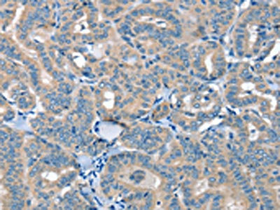
Immunohistochemical staining of human lung shows strong cytoplasmic positivity in pneumocytes and macrophages.

Immunohistochemical staining of human lung shows strong cytoplasmic positivity in pneumocytes and macrophages.
Anti-SELENON Antibody
HPA058076
ApplicationsImmunoCytoChemistry, ImmunoHistoChemistry
Product group Antibodies
ReactivityHuman
TargetSELENON
Overview
- SupplierAtlas Antibodies
- Product NameAnti-SELENON Antibody
- Delivery Days Customer4
- ApplicationsImmunoCytoChemistry, ImmunoHistoChemistry
- CertificationResearch Use Only
- ClonalityPolyclonal
- ConjugateUnconjugated
- Gene ID57190
- Target nameSELENON
- Target descriptionselenoprotein N
- Target synonymsCFTD, CMYO3, CMYP3, MDRS1, RSMD1, RSS, SELN, SEPN1, selenoprotein N, selenoprotein N, 1
- HostRabbit
- IsotypeIgG
- Protein IDQ9NZV5
- Protein NameSelenoprotein N
- Scientific DescriptionRecombinant Protein Epitope Signature Tag (PrEST) antigen sequence
- ReactivityHuman
- Storage Instruction-20°C,2°C to 8°C
- UNSPSC41116161
Datasheet
MSDS
Related products

Product group Antibodies
Anti-SELENON Antibody Picoband(r)A31971-1-CARRIER-FREE
ApplicationsFlow Cytometry, Western Blot, ELISA
ReactivityHuman
TargetSELENON
- SizePrice
Product group Antibodies
Anti-SEPN1 (C-term) Antibody102-21126
ApplicationsWestern Blot, ImmunoHistoChemistry, ImmunoHistoChemistry Paraffin
TargetSELENON
- SizePrice
Product group Antibodies

SELN / SEPN1 AntibodyLS-C749985
ApplicationsWestern Blot
ReactivityHuman, Mouse
TargetSELENON
- SizePrice
Product group Antibodies
Selenon Polyclonal AntibodyCAC11474
ApplicationsWestern Blot, ELISA
ReactivityRat
TargetSELENON
- SizePrice
Product group Antibodies
SEPN1 AntibodyCSB-PA183005
ApplicationsWestern Blot, ELISA, ImmunoHistoChemistry
ReactivityHuman
TargetSELENON
- SizePrice