

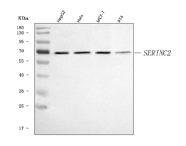
Figure 1. Western blot analysis of SERINC2 using anti-SERINC2 antibody (A14175-2). Electrophoresis was performed on a 5-20% SDS-PAGE gel at 70V (Stacking gel) / 90V (Resolving gel) for 2-3 hours. The sample well of each lane was loaded with 30 ug of sample under reducing conditions. Lane 1: human HepG2 whole cell lysates, Lane 2: human Hela whole cell lysates, Lane 3: human MCF-7 whole cell lysates, Lane 4: human RT4 whole cell lysates. After electrophoresis, proteins were transferred to a nitrocellulose membrane at 150 mA for 50-90 minutes. Blocked the membrane with 5% non-fat milk/TBS for 1.5 hour at RT. The membrane was incubated with rabbit anti-SERINC2 antigen affinity purified polyclonal antibody (Catalog # A14175-2) at 0.5 microg/mL overnight at 4°C, then washed with TBS-0.1%Tween 3 times with 5 minutes each and probed with a goat anti-rabbit IgG-HRP secondary antibody at a dilution of 1:5000 for 1.5 hour at RT. The signal is developed using an Enhanced Chemiluminescent detection (ECL) kit (Catalog # EK1002) with Tanon 5200 system. A specific band was detected for SERINC2 at approximately 65 kDa. The expected band size for SERINC2 is at 51 kDa.
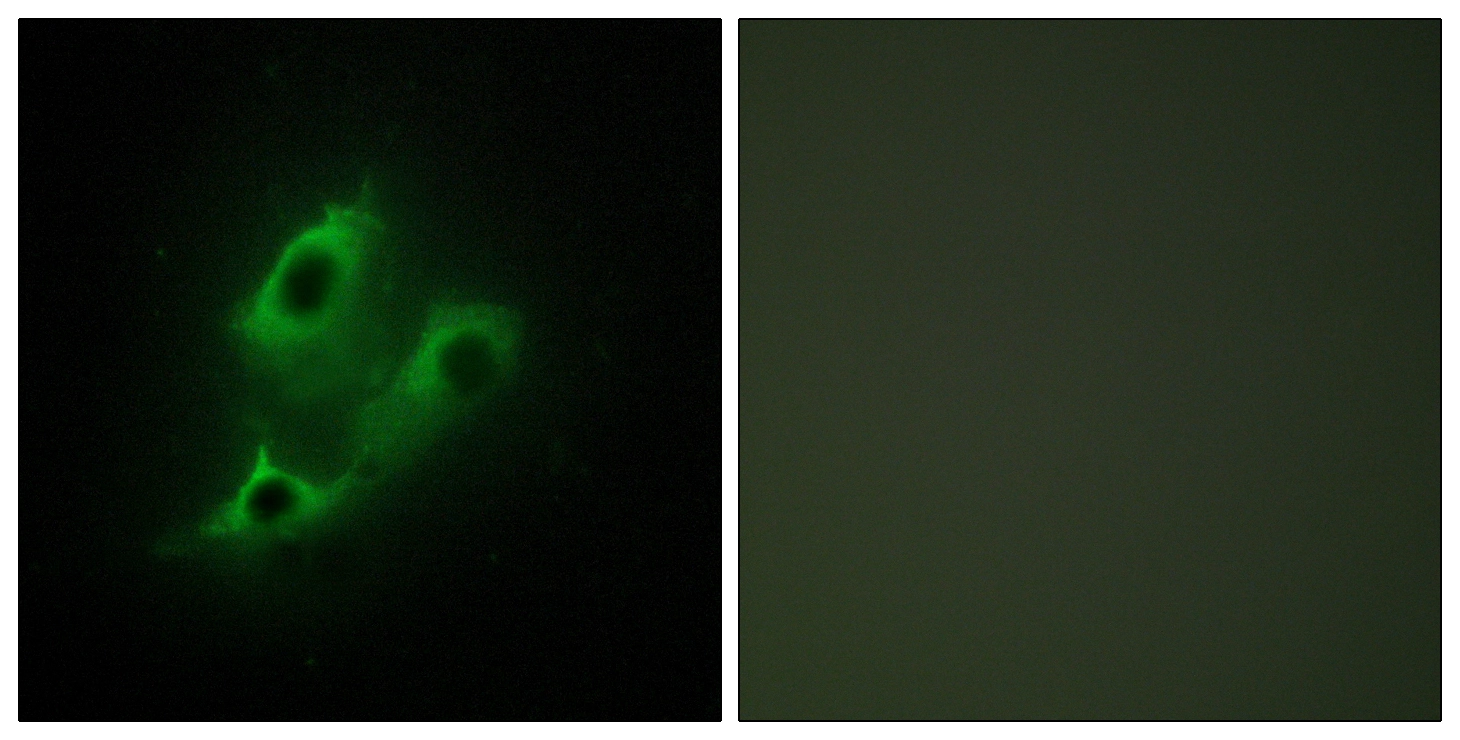
. SERINC2 was detected in an immunocytochemical section of U20S cells. Enzyme antigen retrieval was performed using IHC enzyme antigen retrieval reagent (AR0022) for 15 mins. The cells were blocked with 10% goat serum. And then incubated with 5 microg/mL rabbit anti-SERINC2 Antibody (A14175-2) overnight at 4°C. DyLight®488 Conjugated Goat Anti-Rabbit IgG (BA1127) was used as secondary antibody at 1:500 dilution and incubated for 30 minutes at 37°C. Visualize using a fluorescence microscope and filter sets appropriate for the label used.")
. Overlay histogram showing A431 cells stained with A14175-2 (Blue line). The cells were fixed with 4% paraformaldehyde and blocked with 10% normal goat serum. And then incubated with rabbit anti-SERINC2 Antibody (A14175-2, 1 microg/1x106 cells) for 30 min at 20°C. DyLight®488 conjugated goat anti-rabbit IgG (BA1127, 5-10 microg/1x106 cells) was used as secondary antibody for 30 minutes at 20°C. Isotype control antibody (Green line) was rabbit IgG (1 microg/1x106) used under the same conditions. Unlabelled sample without incubation with primary antibody and secondary antibody (Red line) was used as a blank control.")
Figure 1. Western blot analysis of SERINC2 using anti-SERINC2 antibody (A14175-2). Electrophoresis was performed on a 5-20% SDS-PAGE gel at 70V (Stacking gel) / 90V (Resolving gel) for 2-3 hours. The sample well of each lane was loaded with 30 ug of sample under reducing conditions. Lane 1: human HepG2 whole cell lysates, Lane 2: human Hela whole cell lysates, Lane 3: human MCF-7 whole cell lysates, Lane 4: human RT4 whole cell lysates. After electrophoresis, proteins were transferred to a nitrocellulose membrane at 150 mA for 50-90 minutes. Blocked the membrane with 5% non-fat milk/TBS for 1.5 hour at RT. The membrane was incubated with rabbit anti-SERINC2 antigen affinity purified polyclonal antibody (Catalog # A14175-2) at 0.5 microg/mL overnight at 4°C, then washed with TBS-0.1%Tween 3 times with 5 minutes each and probed with a goat anti-rabbit IgG-HRP secondary antibody at a dilution of 1:5000 for 1.5 hour at RT. The signal is developed using an Enhanced Chemiluminescent detection (ECL) kit (Catalog # EK1002) with Tanon 5200 system. A specific band was detected for SERINC2 at approximately 65 kDa. The expected band size for SERINC2 is at 51 kDa.
Anti-SERINC2 Antibody Picoband(r)
A14175-2-FITC
ApplicationsFlow Cytometry, ImmunoFluorescence, Western Blot, ELISA, ImmunoCytoChemistry
Product group Antibodies
ReactivityHuman
TargetSERINC2
Overview
- SupplierBoster Bio
- Product NameAnti-SERINC2 Antibody Picoband(r)
- Delivery Days Customer9
- ApplicationsFlow Cytometry, ImmunoFluorescence, Western Blot, ELISA, ImmunoCytoChemistry
- CertificationResearch Use Only
- ClonalityPolyclonal
- Concentration500 ug/ml
- ConjugateFITC
- Gene ID347735
- Target nameSERINC2
- Target descriptionserine incorporator 2
- Target synonymsFKSG84, PRO0899, TDE2, TDE2L, serine incorporator 2, tumor differentially expressed protein 2
- HostRabbit
- IsotypeIgG
- Protein IDQ96SA4
- Protein NameSerine incorporator 2
- Scientific DescriptionBoster Bio Anti-SERINC2 Antibody Picoband® catalog # A14175-2. Tested in ELISA, Flow Cytometry, IF, WB, ICC applications. This antibody reacts with Human. The brand Picoband indicates this is a premium antibody that guarantees superior quality, high affinity, and strong signals with minimal background in Western blot applications. Only our best-performing antibodies are designated as Picoband, ensuring unmatched performance.
- ReactivityHuman
- Storage Instruction-20°C,2°C to 8°C
- UNSPSC12352203
Related products
Product group Antibodies
Anti-SERINC2 AntibodyA29390
ApplicationsWestern Blot
ReactivityHuman, Mouse, Rat
- SizePrice
Product group Antibodies

ApplicationsImmunoFluorescence, Western Blot, ELISA, ImmunoHistoChemistry
ReactivityHuman, Mouse, Rat
TargetSERINC2
- SizePrice

Product group Antibodies
Anti-SERINC2 AntibodyHPA005974
ApplicationsWestern Blot, ImmunoHistoChemistry
ReactivityHuman
TargetSERINC2
- SizePrice
Product group Antibodies
SERINC2 AntibodyCSB-PA004253
ApplicationsImmunoFluorescence, Western Blot, ELISA, ImmunoHistoChemistry
ReactivityHuman, Mouse, Rat
TargetSERINC2
- SizePrice
Product group Antibodies
Anti-SERINC2 Antibody Picoband(r)A14175-2-CARRIER-FREE
ApplicationsFlow Cytometry, ImmunoFluorescence, Western Blot, ELISA, ImmunoCytoChemistry
ReactivityHuman
TargetSERINC2
- SizePrice
Product group Antibodies
Anti-SERINC2 (Center) Antibody102-22831
ApplicationsWestern Blot
TargetSERINC2
- SizePrice
Product group Antibodies
SERINC2 antibodyGTX87283
ApplicationsImmunoFluorescence, Western Blot, ImmunoCytoChemistry
ReactivityHuman, Mouse
TargetSERINC2
- SizePrice