


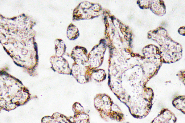
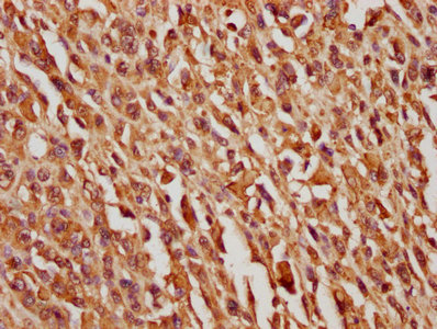
Immunohistochemical staining of human liver shows strong cytoplasmic positivity in hepatocytes.

Immunohistochemical staining of human liver shows strong cytoplasmic positivity in hepatocytes.
Anti-SERPINF1 Antibody
HPA005825
ApplicationsWestern Blot, ImmunoHistoChemistry
Product group Antibodies
ReactivityHuman
TargetSERPINF1
Overview
- SupplierAtlas Antibodies
- Product NameAnti-SERPINF1 Antibody
- Delivery Days Customer4
- ApplicationsWestern Blot, ImmunoHistoChemistry
- CertificationResearch Use Only
- ClonalityPolyclonal
- ConjugateUnconjugated
- Gene ID5176
- Target nameSERPINF1
- Target descriptionserpin family F member 1
- Target synonymsEPC-1, OI12, OI6, PEDF, PIG35, pigment epithelium-derived factor, alpha-2 antiplasmin, cell proliferation-inducing gene 35 protein, serine (or cysteine) proteinase inhibitor, clade F (alpha-2 antiplasmin, pigment epithelium derived factor), member 1, serpin peptidase inhibitor, clade F (alpha-2 antiplasmin, pigment epithelium derived factor), member 1, testis tissue sperm-binding protein Li 70n
- HostRabbit
- IsotypeIgG
- Protein IDP36955
- Protein NamePigment epithelium-derived factor
- Scientific DescriptionRecombinant Protein Epitope Signature Tag (PrEST) antigen sequence
- ReactivityHuman
- Storage Instruction-20°C,2°C to 8°C
- UNSPSC41116161
Datasheet
MSDS
Related products
Product group Antibodies
Anti-PEDF AntibodyA97766
ApplicationsELISA, ImmunoHistoChemistry
ReactivityHuman, Mouse, Rat
- SizePrice
Product group Antibodies
anti-PEDF (human), mAb (rec.) (Serpy-1-4)AG-27B-0014
ApplicationsWestern Blot, ELISA
ReactivityHuman
TargetSERPINF1
- SizePrice
Product group Antibodies
ApplicationsWestern Blot, ELISA
TargetSERPINF1
- SizePrice
Product group Antibodies
PEDF Recombinant Antibody, AbBy Fluor-488 ConjugatedBSM-61731R-BF488
ApplicationsFlow Cytometry, Western Blot
ReactivityHuman
TargetSERPINF1
- SizePrice
Product group Antibodies

Goat anti-SERPINF1 / PEDFEB12223
ApplicationsWestern Blot, ELISA
ReactivityBovine, Canine, Human, Porcine
TargetSERPINF1
- SizePrice
Product group Antibodies
ApplicationsImmunoPrecipitation, Western Blot, ImmunoCytoChemistry, ImmunoHistoChemistry
ReactivityPorcine
TargetSERPINF1
- SizePrice
Product group Antibodies
SERPINF1 AntibodyCSB-PA021084LA01HU
ApplicationsELISA, ImmunoHistoChemistry
ReactivityHuman
TargetSERPINF1
- SizePrice

Product group Antibodies
ApplicationsFlow Cytometry, Western Blot
ReactivityHuman
TargetSERPINF1
- SizePrice
Product group Antibodies

ApplicationsWestern Blot, ELISA
ReactivityHuman
TargetSERPINF1
- SizePrice