


Immunohistochemical staining of human duodenum shows cytoplasmic and membranous positivity in glandular cells.
Immunohistochemical staining of human duodenum shows cytoplasmic and membranous positivity in glandular cells.
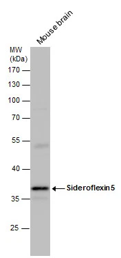
Anti-SFXN5 Antibody
HPA015473
ApplicationsImmunoHistoChemistry
Product group Antibodies
ReactivityHuman
TargetSFXN5
Overview
- SupplierAtlas Antibodies
- Product NameAnti-SFXN5 Antibody
- Delivery Days Customer4
- ApplicationsImmunoHistoChemistry
- CertificationResearch Use Only
- ClonalityPolyclonal
- ConjugateUnconjugated
- Gene ID94097
- Target nameSFXN5
- Target descriptionsideroflexin 5
- Target synonymsBBG-TCC, SLC56A5, sideroflexin-5
- HostRabbit
- IsotypeIgG
- Protein IDQ8TD22
- Protein NameSideroflexin-5
- Scientific DescriptionRecombinant Protein Epitope Signature Tag (PrEST) antigen sequence
- ReactivityHuman
- Storage Instruction-20°C,2°C to 8°C
- UNSPSC41116161
Datasheet
MSDS
Related products

Product group Antibodies
Anti-SFXN5 Antibody Picoband(r)A15698-1-CARRIER-FREE
ApplicationsFlow Cytometry, Western Blot, ELISA, ImmunoHistoChemistry
ReactivityHuman, Mouse, Rat
TargetSFXN5
- SizePrice

Product group Antibodies
SFXN5 AntibodyCSB-PA819464LA01HU
ApplicationsELISA
ReactivityHuman
TargetSFXN5
- SizePrice

Product group Antibodies
Anti-SFXN5 AntibodyHPA056866
ApplicationsImmunoCytoChemistry
ReactivityHuman
TargetSFXN5
- SizePrice
Product group Antibodies
Sideroflexin 5 antibodyGTX120024
ApplicationsWestern Blot
ReactivityHuman, Mouse
TargetSFXN5
- SizePrice
Product group Antibodies

SFXN5 / Sideroflexin 5 AntibodyLS-C399968
ApplicationsELISA
ReactivityHuman
TargetSFXN5
- SizePrice