

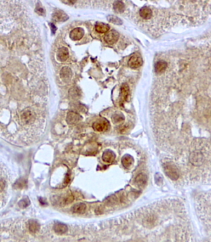
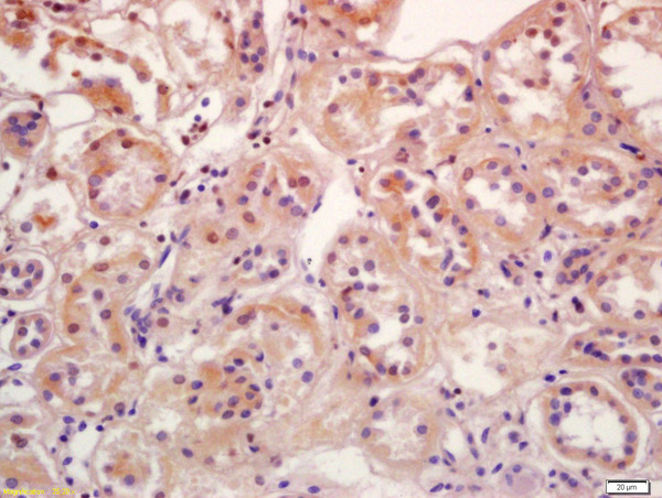
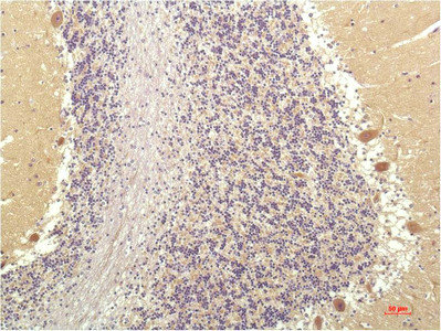
Anti-SGK1 Antibody
A84677
ApplicationsELISA, ImmunoHistoChemistry
Product group Antibodies
ReactivityHuman
Overview
- SupplierAntibodies.com
- Product NameAnti-SGK1 Antibody
- Delivery Days Customer7
- ApplicationsELISA, ImmunoHistoChemistry
- CertificationResearch Use Only
- ClonalityPolyclonal
- Concentration500 ug/ml
- ConjugateUnconjugated
- HostGoat
- IsotypeIgG
- Scientific DescriptionGoat polyclonal antibody to SGK1.
- ReactivityHuman
- UNSPSC12352203
Related products

Product group Antibodies
Anti-SGK1 Antibody Picoband(r)A00673-2-CARRIER-FREE
ApplicationsFlow Cytometry, ImmunoFluorescence, Western Blot, ImmunoCytoChemistry, ImmunoHistoChemistry
ReactivityHuman
TargetSGK1
- SizePrice
Product group Antibodies
Anti-SGK1 Antibody144-01025
ApplicationsImmunoFluorescence, Western Blot, ImmunoHistoChemistry
ReactivityHuman, Mouse, Rat
TargetSGK1
- SizePrice
Product group Antibodies

SGK1 / SGK Antibody (clone 4D12)LS-C770582
ApplicationsImmunoHistoChemistry, ImmunoHistoChemistry Paraffin
ReactivityHuman, Mouse, Rat
TargetSGK1
- SizePrice
Product group Antibodies
ApplicationsFlow Cytometry, ImmunoFluorescence, Western Blot, ELISA, ImmunoCytoChemistry, ImmunoHistoChemistry, ImmunoHistoChemistry Frozen, ImmunoHistoChemistry Paraffin
ReactivityBovine, Canine, Equine, Human, Mouse, Porcine, Rat
TargetSGK1
- SizePrice
Product group Antibodies
SGK1 Monoclonal AntibodyCSB-MA286402
ApplicationsELISA, ImmunoHistoChemistry
ReactivityHuman, Mouse, Rat
TargetSGK1
- SizePrice
Product group Antibodies

Goat anti-SGKEB09396
ApplicationsELISA, ImmunoHistoChemistry
ReactivityBovine, Canine, Human, Mouse, Rat
TargetSGK1
- SizePrice
Product group Antibodies
SGK1 Polyclonal AntibodyCAC13197
ApplicationsImmunoFluorescence, Western Blot, ELISA, ImmunoHistoChemistry
ReactivityMouse
TargetSGK1
- SizePrice

Product group Antibodies
Anti-SGK1 AntibodyHPA051251
ApplicationsImmunoCytoChemistry
ReactivityHuman
TargetSGK1
- SizePrice
Product group Antibodies
SGK1 antibodyGTX107750
ApplicationsWestern Blot
ReactivityHuman, Mouse
TargetSGK1
- SizePrice
Product group Antibodies
Anti-TBX2 AntibodyCAB10250
ApplicationsWestern Blot, ELISA
ReactivityHuman
TargetSGK1
- SizePrice