


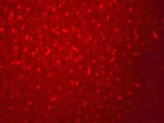
Immunofluorescent staining of human cell line MCF7 shows localization to nucleoplasm.
Immunofluorescent staining of human cell line MCF7 shows localization to nucleoplasm.
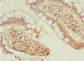
Anti-SIVA1 Antibody
HPA066693
ApplicationsImmunoCytoChemistry
Product group Antibodies
ReactivityHuman
TargetSIVA1
Overview
- SupplierAtlas Antibodies
- Product NameAnti-SIVA1 Antibody
- Delivery Days Customer4
- ApplicationsImmunoCytoChemistry
- CertificationResearch Use Only
- ClonalityPolyclonal
- ConjugateUnconjugated
- Gene ID10572
- Target nameSIVA1
- Target descriptionSIVA1 apoptosis inducing factor
- Target synonymsCD27BP, SIVA, Siva-1, Siva-2, apoptosis regulatory protein Siva, CD27-binding (Siva) protein
- HostRabbit
- IsotypeIgG
- Protein IDO15304
- Protein NameApoptosis regulatory protein Siva
- Scientific DescriptionRecombinant Protein Epitope Signature Tag (PrEST) antigen sequence
- ReactivityHuman
- Storage Instruction-20°C,2°C to 8°C
- UNSPSC41116161
Datasheet
MSDS
Related products
Product group Antibodies
SIVA1 AntibodyCSB-PA021347ESR1HU
ApplicationsELISA, ImmunoHistoChemistry
ReactivityHuman
TargetSIVA1
- SizePrice

Product group Antibodies
Anti-SIVA/SIVA1 Antibody Picoband(r)A05419-1-CARRIER-FREE
ApplicationsFlow Cytometry, Western Blot, ELISA
ReactivityHuman
TargetSIVA1
- SizePrice

Product group Antibodies
Anti-SIVA1 AntibodyHPA065398
ApplicationsImmunoCytoChemistry
ReactivityHuman
TargetSIVA1
- SizePrice
Product group Antibodies
Anti-SIVA1 AntibodyHPA065398
ApplicationsImmunoCytoChemistry
ReactivityHuman
TargetSIVA1
- SizePrice
Product group Antibodies

SIVA1 / SIVA Antibody (aa1-110)LS-C370333
ApplicationsWestern Blot, ELISA
ReactivityHuman
TargetSIVA1
- SizePrice
Product group Antibodies
ApplicationsImmunoPrecipitation, Western Blot, ImmunoCytoChemistry, ImmunoHistoChemistry
TargetSIVA1
- SizePrice
Product group Antibodies
SIVA antibodyGTX32046
ApplicationsWestern Blot, ELISA, ImmunoHistoChemistry, ImmunoHistoChemistry Paraffin
ReactivityHuman, Mouse
TargetSIVA1
- SizePrice