


Immunohistochemical staining of human testis shows strong membranous positivity in cells in seminiferous ducts.
Immunohistochemical staining of human testis shows strong membranous positivity in cells in seminiferous ducts.
Anti-SLC7A5 Antibody
HPA052673
ApplicationsImmunoHistoChemistry
Product group Antibodies
ReactivityHuman
TargetSLC7A5
Overview
- SupplierAtlas Antibodies
- Product NameAnti-SLC7A5 Antibody
- Delivery Days Customer4
- ApplicationsImmunoHistoChemistry
- CertificationResearch Use Only
- ClonalityPolyclonal
- ConjugateUnconjugated
- Gene ID8140
- Target nameSLC7A5
- Target descriptionsolute carrier family 7 member 5
- Target synonyms4F2LC, CD98, D16S469E, E16, LAT1, MPE16, large neutral amino acids transporter small subunit 1, 4F2 light chain, CD98 light chain, L-type amino acid transporter 1, integral membrane protein E16, sodium-independent neutral amino acid transporter LAT1, solute carrier family 7 (amino acid transporter light chain, L system), member 5, solute carrier family 7 (cationic amino acid transporter, y+ system), member 5
- HostRabbit
- IsotypeIgG
- Protein IDQ01650
- Protein NameLarge neutral amino acids transporter small subunit 1
- Scientific DescriptionRecombinant Protein Epitope Signature Tag (PrEST) antigen sequence
- ReactivityHuman
- Storage Instruction-20°C,2°C to 8°C
- UNSPSC41116161
Datasheet
MSDS
Related products
Product group Antibodies
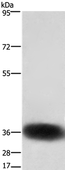
Anti-SLC7A5 Antibody144-02833
ApplicationsWestern Blot
ReactivityHuman, Mouse, Rat
TargetSLC7A5
- SizePrice
Product group Antibodies
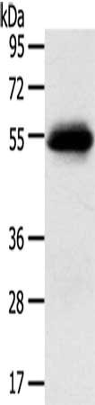
Anti-SLC7A5 AntibodyA42423
ApplicationsWestern Blot
ReactivityHuman, Mouse
- SizePrice
Product group Antibodies

SLC7A5 / CD98 Light Chain AntibodyLS-C667860
ApplicationsWestern Blot
ReactivityHuman
TargetSLC7A5
- SizePrice
Product group Antibodies

Anti-CD98 [EP3-1]Ab00898-1.1
ApplicationsImmunoFluorescence, ImmunoPrecipitation, Other Application
ReactivityHuman
TargetSLC7A5
- SizePrice
Product group Antibodies
References
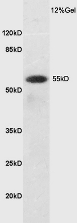
SLC7A5 Polyclonal AntibodyBS-10125R
ApplicationsImmunoFluorescence, Western Blot, ELISA, ImmunoCytoChemistry, ImmunoHistoChemistry, ImmunoHistoChemistry Paraffin
ReactivityHuman, Mouse, Rat
TargetSLC7A5
- SizePrice
Product group Antibodies

Goat anti-LAT1 / SLC7A5EB09262
ApplicationsWestern Blot, ELISA
ReactivityCanine, Human
TargetSLC7A5
- SizePrice
Product group Antibodies
SLC7A5 AntibodyCSB-PA057466
ApplicationsWestern Blot, ELISA
ReactivityHuman
TargetSLC7A5
- SizePrice

Product group Antibodies
Anti-SLC7A5 Antibody (N-Term)M01016-1
ApplicationsImmunoFluorescence, Western Blot
ReactivityHuman
TargetSLC7A5
- SizePrice